A Spine MRI for back pain is the definitive way to see exactly what is happening inside your body when symptoms refuse to settle. Up to 80% of adults will experience lower back pain at some point in their lives. In the vast majority of cases, the pain is muscular or mechanical, resolving within a few weeks with rest and physiotherapy. However, when the pain is unrelenting or shoots down the leg, patients often wonder if they need a scan. Dr Prashant Sankaye, expert MSK Radiologist at London Sports & Rheumatology Imaging (LSRI), outlines exactly when this high-resolution imaging becomes essential.
What Does a Spine MRI for Back Pain Show?
Unlike a traditional X-ray, which only evaluates the bony vertebrae, an MRI offers an exquisite, highly detailed look at the soft tissues. It clearly illuminates the spinal cord, the exiting nerve roots, and the intervertebral discs. Today, a Spine MRI for back pain is universally recognised as the gold standard for diagnosing complex spinal pathologies that other imaging methods simply cannot detect.
7 Crucial Signs You Need a Spine MRI for Back Pain
Dr Prashant Sankaye generally advises observing mild symptoms for 4 to 6 weeks alongside conservative treatment, such as targeted physiotherapy. However, you should not wait to book a Spine MRI for back pain if you experience any of these major clinical “red flags”:
-
Severe Sciatica: Intense, shooting pain radiating past the knee, into the calf or foot.
-
Neurological Symptoms: Numbness, tingling, or sudden weakness in the foot (such as foot drop).
-
History of Trauma: A recent significant fall, sports injury, or road traffic accident.
-
Unexplained Weight Loss: When coupled with back pain, this can occasionally indicate more systemic or underlying medical issues.
-
Night Sweats and Night Pain: Pain that is worse at night and disrupts your sleep continuously.
-
Lack of Improvement: Pain that remains severe and unchanging despite 6 weeks of dedicated physiotherapy.
-
Loss of Bladder or Bowel Control: This is a medical emergency known as Cauda Equina Syndrome. It requires an immediate hospital visit and an urgent scan.
Note: For standard acute pain without these red flags, NICE guidelines on sciatica recommend waiting to see if symptoms resolve before seeking specialist imaging.
Common Diagnoses Found on Your Scan
If your imaging is performed at LSRI, Dr Prashant Sankaye will carefully evaluate the images for the following common culprits:
-
Herniated (Slipped) Disc: This occurs when the soft inner core of a spinal disc pushes through its tough exterior. It can physically impinge on a nearby nerve root, causing intense sciatic pain.
-
Spinal Stenosis: This is a narrowing of the spinal canal. It is typically due to age-related osteoarthritis and the thickening of spinal ligaments, which slowly compresses the spinal nerves.
-
Spondylolisthesis: A mechanical condition where one vertebra slips forward over the bone directly beneath it, causing instability and nerve irritation.
What to Expect During Your Appointment
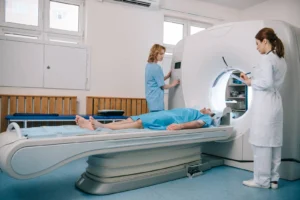
Having a Spine MRI for back pain is a completely painless and non-invasive procedure. At LSRI, our state-of-the-art 3T MRI scanners provide a spacious and comfortable environment. The scan typically takes between 20 to 30 minutes, during which you simply lie still. Because MRI uses powerful magnetic fields instead of ionising radiation, it is extremely safe for patients.
The LSRI Diagnostic Difference
At London Sports & Rheumatology Imaging, we ensure your scan is reported with your specific clinical symptoms in mind. Avoid unnecessary anxiety by having your imaging interpreted directly by a sub-specialist MSK Radiologist.
Getting a highly detailed Spine MRI for back pain provides the clarity you and your treating physician need to map out an effective, long-lasting treatment plan. If you are struggling with unresolved symptoms and need answers, get in touch today to arrange a high-resolution 3T MRI scan with Dr Prashant Sankaye’s expert reporting.
About the Author: Dr Prashant Sankaye, Consultant Musculoskeletal specialist and Radiologist, MBBS, MS, FCPS, MRCS, CCBST, FRCR, PGCE(Med), FHEA, PGDip Sports and Exercise Medicine
Dr Prashant Sankaye is a highly respected Consultant MSK Radiologist and the Clinical Director of London Sports & Rheumatology Imaging (LSRI). With over a decade of sub-specialty experience, he is a recognized expert in advanced diagnostic imaging (Ultrasound & 3T MRI) and precision ultrasound-guided therapeutic injections. His authoritative approach ensures patients avoid surgery where possible and receive the highest standard of orthopaedic, rheumatological, and sports medicine care.