Written by: LSRI Editorial Team
Medically Reviewed by: Dr Prashant Sankaye, Consultant MSK Radiologist — MBBS, MS, FCPS, MRCS, CCBST, FRCR, PGCE(Med), FHEA, PGDip Sports and Exercise Medicine
Last Updated: October 14, 2026
Read Time: 6 Minutes
If you have recently undergone an MRI scan for joint or muscle pain, reading the radiologist’s mri scan results can feel like learning a foreign language. Terms like “effusion,” “meniscal tear,” or “tendinosis” stand between you and understanding your diagnosis. At London Sports & Rheumatology Imaging (LSRI), Dr Prashant Sankaye, a leading Consultant MSK Radiologist, believes patient education is the cornerstone of effective recovery.
Why Are MRI Scans So Detailed?
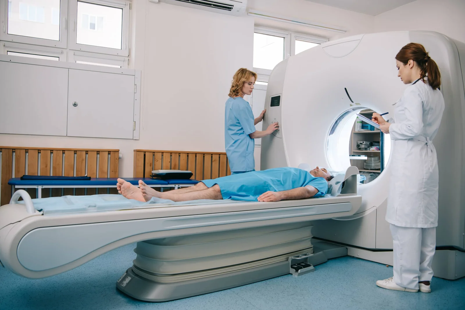
An MRI scanner uses powerful magnetic fields to create highly detailed, cross-sectional images of your internal structures. Unlike X-rays, which mainly show bone, MRI reveals the soft tissues—cartilage, ligaments, tendons, and muscles—where the vast majority of sports and degenerative injuries occur.
Common Terms Decoded by Dr Prashant Sankaye
- Joint Effusion: This simply means “swelling.” It indicates increased synovial fluid in a joint, usually a reaction to inflammation or injury.
- Tendinopathy / Tendinosis: This refers to chronic degeneration or micro-tearing in a tendon (such as the Achilles or Patellar tendon), rather than acute inflammation (tendinitis).
- Meniscal Tear: A common knee injury involving the shock-absorbing cartilage. The MRI will detail whether it is a radial, horizontal, or complex tear, which dictates whether conservative treatment or surgery is needed.
- Osteoarthritis / Chondromalacia: These terms describe the thinning, softening, or loss of the articular cartilage protecting the ends of your bones.
When an MRI Leads to Action
According to Dr Prashant Sankaye, “An MRI shouldn’t just be a picture; it should be a roadmap for your treatment.” Once the exact source of the pain is pinpointed, targeted interventions such as ultrasound-guided injections can deliver powerful anti-inflammatory medication or regenerative biologics directly to the site of the injury.
“Do not panic if your MRI report lists multiple findings. Degenerative changes are often a normal part of aging. The key is treating the patient, not just the scan.” — Dr Prashant Sankaye
Get Your Scan Reviewed at LSRI
Are you looking for a Second Opinion on a recent MRI scan? Dr Prashant Sankaye provides expert, sub-specialist reviews of MRI scans and reports. Book a consultation at LSRI today.
What Is an MRI Scan Report?
After your MRI scan, a Consultant Radiologist — in our case, Dr Prashant Sankaye — analyses the images and produces a formal written report. This report describes everything observed on the scan: the structures examined, any abnormal findings, and their likely clinical significance. Understanding your MRI scan results begins with recognising that the report is written for clinicians, using anatomical and radiological terminology — but with a little guidance, most patients can understand the key findings.
Common Terms in MRI Scan Reports — Explained
Below are the most frequently encountered terms in musculoskeletal MRI reports, translated into plain English:
Terms Related to Joints
- Effusion — excess fluid within the joint space, indicating inflammation or injury
- Osteophyte — bony spur, typically associated with osteoarthritis and joint degeneration
- Subchondral oedema — bone marrow swelling beneath the cartilage, often indicating bone stress or early arthritis
- Chondromalacia — softening and breakdown of cartilage, commonly seen under the kneecap
- Synovitis — inflammation of the joint lining, common in inflammatory arthritis
Terms Related to Tendons and Ligaments
- Tendinopathy / tendinosis — degenerative change within a tendon, presenting as signal change on MRI
- Partial tear — incomplete disruption of a tendon or ligament fibres
- Full-thickness tear — complete rupture through the entire thickness of a tendon or ligament
- High signal / increased signal — an area that appears bright on certain MRI sequences, usually indicating fluid, inflammation, or tear
Terms Related to Muscles
- Myositis — muscle inflammation
- Intramuscular haematoma — a bruise within the muscle tissue
- Muscle oedema — fluid accumulation within muscle, indicating strain or overuse
Terms Related to Bone
- Bone marrow oedema — swelling within the bone, indicating stress, injury, or inflammation
- Stress fracture — a crack in the bone caused by repetitive loading
- Avascular necrosis (AVN) — death of bone tissue due to loss of blood supply
- Sclerosis — abnormal hardening of bone tissue
How to Read Your MRI Report: A Step-by-Step Approach
When you receive your MRI scan results, follow this structured approach:
- Read the clinical indication — this describes why the scan was requested and what was being investigated
- Read the findings section — this is the detailed description of what was observed on each sequence
- Read the conclusion or impression — this is the radiologist’s summary, highlighting the most important findings and their likely clinical significance
- Identify any urgent findings — urgent findings requiring immediate action are usually flagged clearly, for example “recommend urgent orthopaedic review”
The Royal College of Radiologists publishes standards for how MRI reports should be structured and communicated to patients and referring clinicians.
What If Your MRI Scan Is Normal?
A normal MRI report can be reassuring — it rules out serious structural pathology. However, a normal MRI does not necessarily mean there is no pain or clinical problem. Some conditions, such as fibromyalgia or early inflammatory arthritis, may not produce visible changes on MRI. Your clinician will interpret the normal result in the context of your symptoms and examination findings.
What If Your MRI Shows an Unexpected Finding?
Radiologists sometimes identify incidental findings — abnormalities not related to the original clinical question. Common incidental findings in musculoskeletal MRI include small cysts, mild degenerative changes, or minor signal changes. Dr Sankaye always contextualises these findings, indicating whether they require follow-up or are of no clinical significance.
Discussing Your MRI Results with Dr Sankaye
At LSRI, we believe patients deserve to fully understand their imaging results. Our reports are written with clarity in mind, and Dr Sankaye is available to discuss your findings in a follow-up consultation. We can explain exactly what your MRI shows, what it means for your treatment options, and what the recommended next steps are.
According to Radiopaedia, one of the world’s leading radiology education resources, patient-centred reporting is increasingly recognised as best practice in modern clinical radiology.
Book Your MRI Scan and Expert Reporting in London
Whether you have already had a scan and want a second opinion, or you need a new MRI scan for an ongoing musculoskeletal issue, LSRI offers expert imaging and interpretation services in central London. Contact us today to book your appointment.
Frequently Asked Questions
How long does it take to get MRI results at LSRI?
Your formal written report is available within 24 hours of your scan. To go through the results with a specialist, choose MRI with a consultation (from £425).
Can I request a copy of my MRI images?
Yes. You are entitled to a copy of your scan images. Please ask our team when booking.
What if I disagree with the findings on my MRI report?
You can request a second opinion from another consultant radiologist. Dr Sankaye provides independent second opinion reporting for patients with complex or disputed findings.