Written by: LSRI Editorial Team
Medically Reviewed by: Dr Prashant Sankaye, Consultant MSK Radiologist — MBBS, MS, FCPS, MRCS, CCBST, FRCR, PGCE(Med), FHEA, PGDip Sports and Exercise Medicine
Last Updated: October 11, 2026
Read Time: 7 Minutes
A Spine MRI for back pain is the definitive way to see exactly what is happening inside your body when symptoms refuse to settle. Up to 80% of adults will experience lower back pain at some point in their lives. In the vast majority of cases, the pain is muscular or mechanical, resolving within a few weeks with rest and physiotherapy. However, when the pain is unrelenting or shoots down the leg, patients often wonder if they need a scan. Dr Prashant Sankaye, expert MSK Radiologist at London Sports & Rheumatology Imaging (LSRI), outlines exactly when this high-resolution imaging becomes essential.
What Does a Spine MRI for Back Pain Show?
Unlike a traditional X-ray, which only evaluates the bony vertebrae, an MRI offers an exquisite, highly detailed look at the soft tissues. It clearly illuminates the spinal cord, the exiting nerve roots, and the intervertebral discs. Today, a Spine MRI for back pain is universally recognised as the gold standard for diagnosing complex spinal pathologies that other imaging methods simply cannot detect.
7 Crucial Signs You Need a Spine MRI for Back Pain
Dr Prashant Sankaye generally advises observing mild symptoms for 4 to 6 weeks alongside conservative treatment, such as targeted physiotherapy. However, you should not wait to book a Spine MRI for back pain if you experience any of these major clinical “red flags”:
-
Severe Sciatica: Intense, shooting pain radiating past the knee, into the calf or foot.
-
Neurological Symptoms: Numbness, tingling, or sudden weakness in the foot (such as foot drop).
-
History of Trauma: A recent significant fall, sports injury, or road traffic accident.
-
Unexplained Weight Loss: When coupled with back pain, this can occasionally indicate more systemic or underlying medical issues.
-
Night Sweats and Night Pain: Pain that is worse at night and disrupts your sleep continuously.
-
Lack of Improvement: Pain that remains severe and unchanging despite 6 weeks of dedicated physiotherapy.
-
Loss of Bladder or Bowel Control: This is a medical emergency known as Cauda Equina Syndrome. It requires an immediate hospital visit and an urgent scan.
Note: For standard acute pain without these red flags, it is often reasonable to wait and see if symptoms settle before seeking specialist imaging.
Common Diagnoses Found on Your Scan
If your imaging is performed at LSRI, Dr Prashant Sankaye will carefully evaluate the images for the following common culprits:
-
Herniated (Slipped) Disc: This occurs when the soft inner core of a spinal disc pushes through its tough exterior. It can physically impinge on a nearby nerve root, causing intense sciatic pain.
-
Spinal Stenosis: This is a narrowing of the spinal canal. It is typically due to age-related osteoarthritis and the thickening of spinal ligaments, which slowly compresses the spinal nerves.
-
Spondylolisthesis: A mechanical condition where one vertebra slips forward over the bone directly beneath it, causing instability and nerve irritation.
What to Expect During Your Appointment
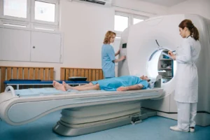
Having a Spine MRI for back pain is a completely painless and non-invasive procedure. At LSRI, spine MRI is carried out at our partner imaging centres, on a 1.5T or 3T scanner. The scan typically takes between 20 to 30 minutes, during which you simply lie still. Because MRI uses powerful magnetic fields instead of ionising radiation, it is very safe for most people, but some implants and devices, such as certain pacemakers, must be checked first, so you will be asked a set of safety questions before your scan.
The LSRI Diagnostic Difference
At London Sports & Rheumatology Imaging, we ensure your scan is reported with your specific clinical symptoms in mind. Avoid unnecessary anxiety by having your imaging interpreted directly by a sub-specialist MSK Radiologist.
Getting a highly detailed Spine MRI for back pain provides the clarity you and your treating physician need to map out an effective, long-lasting treatment plan. If you are struggling with unresolved symptoms and need answers, get in touch today to arrange an MRI scan reported by Dr Prashant Sankaye, Consultant MSK Radiologist.
What Does a Spine MRI Show That Other Scans Miss?
X-rays visualise bone only. CT scans provide excellent bony detail but use ionising radiation and are less sensitive for soft tissue pathology. A spine MRI, by contrast, simultaneously images the vertebral bodies, intervertebral discs, spinal cord, nerve roots, facet joints, paraspinal muscles, and ligaments — all in a single, radiation-free examination.
This makes spine MRI the definitive investigation for:
- Disc herniation — prolapsed disc material pressing on nerve roots, causing sciatica or referred arm pain
- Spinal stenosis — narrowing of the spinal canal causing neurogenic claudication (leg pain on walking)
- Facet joint arthropathy — degenerative changes in the posterior joints causing localised back pain
- Spondylolisthesis — slippage of one vertebra over another, visible on dynamic or standard MRI
- Cauda equina syndrome — compression of the nerve bundle at the base of the spine, a medical emergency
- Vertebral fractures — particularly insufficiency fractures in osteoporotic patients
- Inflammatory spinal disease — including ankylosing spondylitis and sacroiliitis, where bone marrow oedema is the earliest sign
- Spinal tumours and metastases — including bone metastases from prostate, breast, lung, and renal cancers
- Disc infection (discitis) — infection of the intervertebral disc and adjacent vertebral bodies
Which strength of MRI scanner is used for the spine?
The strength of an MRI scanner is measured in tesla (T). LSRI arranges spine MRI at partner imaging centres with 1.5T and 3T scanners. A 1.5T scanner gives diagnostic images for the large majority of spinal problems, and 3T is available when it adds value. The scan protocol and the experience of the radiologist reporting the scan matter as much as the field strength.
MRI is the preferred imaging test when nerve root or cord compression is suspected clinically.
The 7 Crucial Signs You Need a Spine MRI
1. Sciatica Lasting More Than 6 Weeks
Sciatic pain radiating from the lower back into the buttock, thigh, calf, or foot — especially when associated with numbness, tingling, or weakness — is a strong indicator for MRI. While most acute sciatica resolves conservatively, persistent symptoms beyond 4–6 weeks warrant imaging to identify the causative pathology and guide treatment.
2. Red Flag Back Pain Symptoms
Certain symptoms require urgent MRI referral: bladder or bowel dysfunction alongside back pain, saddle anaesthesia (numbness around the groin and inner thighs), rapidly progressive leg weakness, or bilateral leg symptoms. These may indicate cauda equina syndrome — a surgical emergency.
3. Back Pain Following Trauma
Back pain after a fall, road traffic accident, or sporting injury requires imaging to exclude vertebral fracture, ligamentous injury, or spinal cord contusion.
4. Pain Unresponsive to Conservative Treatment
If back pain has persisted despite 6–12 weeks of physiotherapy, analgesia, and lifestyle modification, MRI provides the structural diagnosis needed to direct more targeted treatment.
5. Suspected Inflammatory Spinal Disease
Young patients with morning stiffness, sacroiliac pain, and back pain that improves with exercise may have ankylosing spondylitis. MRI of the sacroiliac joints and spine is essential for early diagnosis before X-ray changes appear.
6. History of Cancer
Any patient with a history of malignancy who presents with new back pain requires urgent spine MRI to exclude vertebral metastases or epidural cord compression.
7. Night Pain or Systemic Symptoms
Back pain that is constant, severe at night, associated with unexplained weight loss, fever, or night sweats should be investigated urgently with MRI and laboratory tests to exclude infection (discitis, epidural abscess) or malignancy.
Booking a Spine MRI at LSRI London
At LSRI, spine MRI scans are available with same-day or next-day appointments. All scans are reported by Dr Prashant Sankaye, with written reports available within 24 hours. We accept self-referrals, GP referrals, and consultant referrals. Book your spine MRI today.
For further reading on spinal conditions and MRI, the Radiopaedia spinal MRI guide is an excellent clinical reference.