✍️ Written by: LSRI Editorial Team
🩺 Medically Reviewed by: Dr Prashant Sankaye, Consultant Musculoskeletal specialist and Radiologist, MBBS, MS, FCPS, MRCS, CCBST, FRCR, PGCE(Med), FHEA, PGDip Sports and Exercise Medicine
📅 Last Updated: March 17, 2026
⏱️ Read Time: 2 Minutes
You have a deep, aching pain in your shoulder, hip, or knee. Is it arthritis, or is it a tendon issue? Clinically, the symptoms of these two vastly different conditions often overlap—presenting as stiffness, weakness, and pain during movement. Dr Prashant Sankaye, Consultant MSK Radiologist at London Sports & Rheumatology Imaging, explains how advanced diagnostic imaging is the ultimate tool for cutting through the confusion.
Understanding the Anatomy
Arthritis (Osteoarthritis): This is an intra-articular (inside the joint) problem. It involves the progressive wearing away of the smooth articular cartilage that caps the ends of your bones. As the cartilage degrades, the bones rub together, causing pain, bone spurs, and deep joint inflammation.
Tendonitis/Tendinopathy: This is an extra-articular (outside the joint) problem. Tendons are the thick fibrous cords that attach your muscles to your bones. When overused or strained, they can develop micro-tears, inflammation, and thickening.
How Do We Tell the Difference?
While a physical examination provides clues, only imaging can offer definitive proof. Dr Prashant Sankaye utilises both Ultrasound and MRI depending on the joint location.
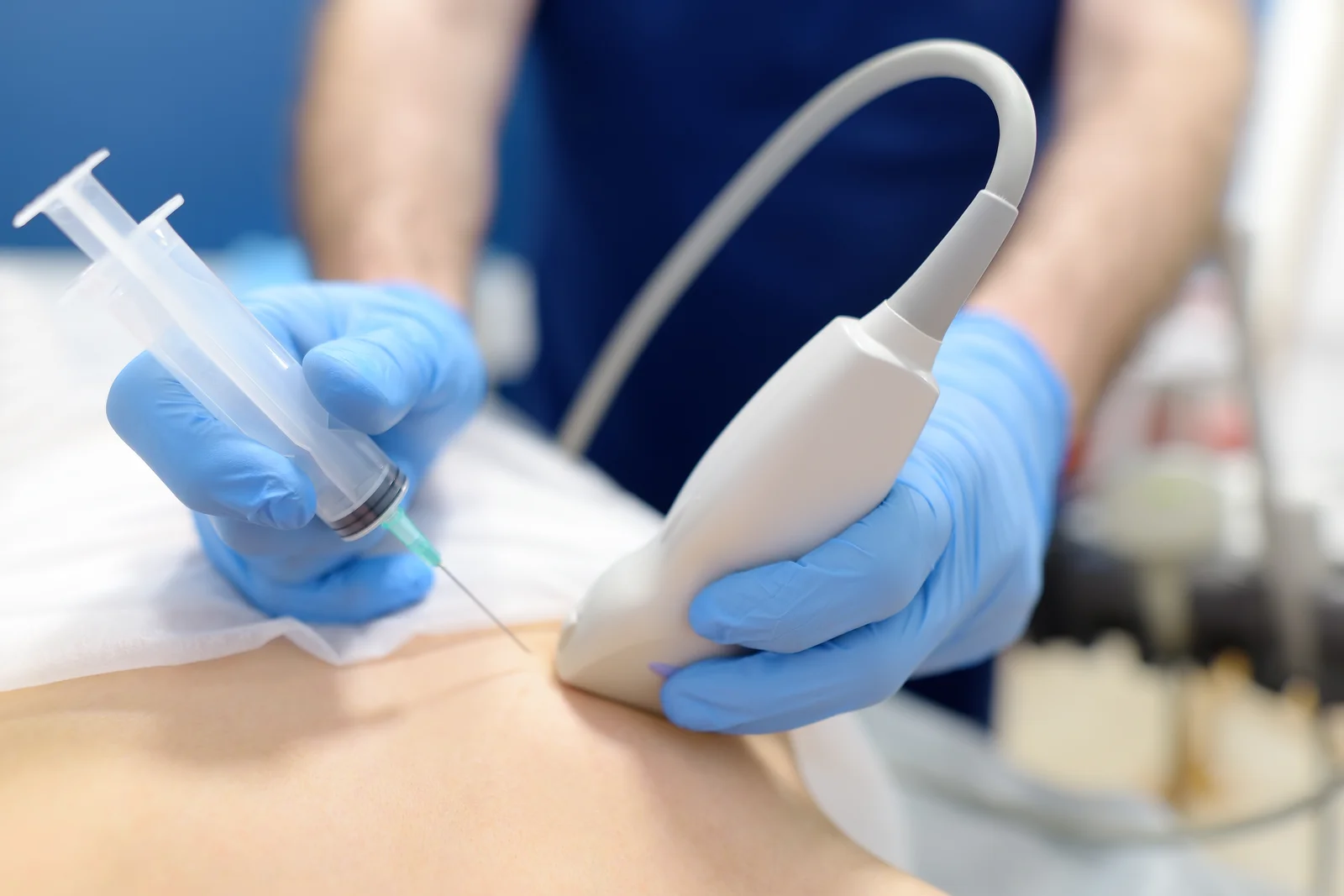
Diagnostic Ultrasound
An Ultrasound scan is exceptional for evaluating tendons in real-time. Dr Sankaye can ask you to move your shoulder or knee while scanning, actively watching how the tendon glides. Ultrasound can easily identify thickening, tears, and active inflammation within the tendon. It can also detect fluid build-up inside the joint, aiming us toward an arthritis diagnosis.
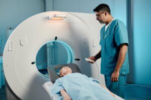
3T MRI
An MRI scan provides a comprehensive 3D view of both the inside and outside of the joint. It is unrivalled in its ability to evaluate the exact thickness and quality of the articular cartilage (key for diagnosing arthritis severity) while simultaneously clearly showing the health of all surrounding tendons and ligaments.
Why Diagnostic Certainty Matters
Treatment for tendonitis differs substantially from arthritis. Tendon issues may require targeted physiotherapy or PRP injections to stimulate healing. Arthritis may require viscosupplements like Arthrosamid or Hyaluronic acid to lubricate the joint.
“Guessing the source of musculoskeletal pain leads to failed treatments and frustrated patients. Imaging provides absolute clarity.” — Dr Prashant Sankaye
Start Your Recovery Today
If you are experiencing chronic joint pain and standard treatments aren’t working, an accurate diagnosis is your missing link. Contact LSRI to book a comprehensive diagnostic scan with Dr Prashant Sankaye.
About the Author: Dr Prashant Sankaye, Consultant Musculoskeletal specialist and Radiologist, MBBS, MS, FCPS, MRCS, CCBST, FRCR, PGCE(Med), FHEA, PGDip Sports and Exercise Medicine
Dr Prashant Sankaye is a highly respected Consultant MSK Radiologist and the Clinical Director of London Sports & Rheumatology Imaging (LSRI). With over a decade of sub-specialty experience, he is a recognized expert in advanced diagnostic imaging (Ultrasound & 3T MRI) and precision ultrasound-guided therapeutic injections. His authoritative approach ensures patients avoid surgery where possible and receive the highest standard of orthopaedic, rheumatological, and sports medicine care.