✍️ Written by: LSRI Editorial Team
🩺 Medically Reviewed by: Dr Prashant Sankaye, Consultant Musculoskeletal specialist and Radiologist, MBBS, MS, FCPS, MRCS, CCBST, FRCR, PGCE(Med), FHEA, PGDip Sports and Exercise Medicine
📅 Last Updated: March 12, 2026
⏱️ Read Time: 6 Minutes
Sports injuries are an unfortunate reality for athletes and active individuals, ranging from minor sprains to more complex conditions that require precise diagnosis and treatment. Accurate assessment of these injuries is vital not only for effective treatment but also for reducing downtime and preventing long-term complications. This is where advanced imaging techniques, such as MRI, ultrasound, and X-ray, play a transformative role.
In this blog post, we will explore the importance of advanced imaging in diagnosing sports injuries, the types of imaging available, how each technique works, and the benefits they offer to athletes and healthcare providers alike.
The Importance of Accurate Diagnosis in Sports Injuries
Sports injuries often involve complex musculoskeletal structures such as ligaments, tendons, cartilage, and bone. Accurate diagnosis is crucial for several reasons:
- Pinpointing the Cause of Pain: Identifying the exact structure affected is key to developing a tailored treatment plan.
- Guiding Treatment: Whether it’s rehabilitation, medication, or surgery, advanced imaging helps determine the best course of action.
- Preventing Further Injury: Misdiagnosis or delayed treatment can lead to worsening injuries or chronic issues.
- Improving Recovery Time: Precise imaging allows for targeted interventions, which can speed up recovery.
Traditional diagnostic methods, such as physical exams and patient history, are still important but are often insufficient for fully understanding the extent of an injury. This is where advanced imaging steps in, offering detailed insights that cannot be obtained otherwise.
Types of Advanced Imaging Techniques
Advanced imaging techniques revolutionise the diagnosis and management of sports injuries by providing detailed visuals of the affected areas. The primary modalities include:
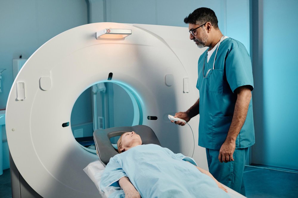
- Magnetic Resonance Imaging (MRI)
MRI is one of the most powerful tools for diagnosing sports injuries. Using strong magnetic fields and radio waves, it creates detailed images of soft tissues, including muscles, ligaments, and tendons.
- Best For: Diagnosing ligament tears (e.g., ACL), meniscal injuries, muscle strains, cartilage damage, and stress fractures.
- Advantages:
- Non-invasive and radiation-free.
- Excellent for detecting soft tissue abnormalities.
- Provides a multi-dimensional view of the affected area.
- Limitations:
- Expensive compared to other modalities.
- Not suitable for patients with certain implants or claustrophobia.
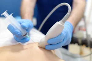
- Ultrasound Imaging
Ultrasound uses high-frequency sound waves to create real-time images of muscles, tendons, and joints. It is often used for dynamic assessments, where the injured area is observed during movement.
- Best For: Identifying tendon tears, muscle injuries, ligament damage, and bursitis.
- Advantages:
- Real-time imaging allows for functional assessment.
- Portable and cost-effective.
- Useful for guiding injections or aspiration procedures.
- Limitations:
- Operator-dependent, requiring skilled practitioners for accurate interpretation.
- Limited penetration depth, less effective for deep structures.
- X-Ray
X-rays are one of the most common imaging tools used in sports medicine. They are excellent for visualising bone structures and detecting fractures.
- Best For: Identifying fractures, dislocations, and bone deformities.
- Advantages:
- Widely available and quick to perform.
- Useful as an initial diagnostic tool.
- Limitations:
- Ineffective for soft tissue injuries.
- Involves exposure to ionising radiation.
- CT Scans
Computed Tomography (CT) scans use a combination of X-rays and computer processing to create detailed cross-sectional images of the body. They are particularly useful for complex bone injuries.
- Best For: Visualising complex fractures and joint damage.
- Advantages:
- Highly detailed images of bone and joint structures.
- Can be used to create 3D reconstructions.
- Limitations:
- Higher radiation dose than standard X-rays.
- Limited soft tissue visualisation.
How Advanced Imaging Works in Diagnosing Sports Injuries
Initial Assessment and Referral
The process typically begins with a physical examination by a sports physician or physiotherapist. If the injury is suspected to involve deeper structures or does not respond to initial treatments, advanced imaging is recommended.
Choosing the Right Imaging Modality
The choice of imaging depends on the suspected injury:
- MRI for soft tissue injuries such as ligament or cartilage tears.
- Ultrasound for tendon and muscle assessments.
- X-rays or CT scans for fractures and joint dislocations.
Capturing the Images
During the imaging session, the patient is positioned to allow clear visualisation of the affected area. For example:
- An MRI scan may involve lying still in a scanner for 20-40 minutes.
- Ultrasound is often performed dynamically, with the patient moving the joint or muscle being examined.
Interpreting the Results
Once the images are captured, a radiologist or imaging specialist reviews them to identify abnormalities. These results are then shared with the referring physician to guide treatment decisions.
The Benefits of Advanced Imaging for Sports Injuries
Advanced imaging techniques offer several advantages over traditional diagnostic methods:
- Detailed Visualisation
Imaging provides high-resolution, detailed views of internal structures, allowing for precise identification of injuries that may not be visible through physical examination. - Enhanced Treatment Planning
With a clear understanding of the injury, healthcare providers can develop targeted treatment plans, whether that involves surgery, rehabilitation, or injections. - Reduced Recovery Times
Accurate diagnosis helps avoid trial-and-error treatments, allowing athletes to begin the correct intervention sooner. - Minimally Invasive Procedures
Imaging techniques like ultrasound are often used to guide minimally invasive treatments, such as PRP injections or aspirations. - Improved Patient Confidence
Clear, visual evidence of an injury helps patients better understand their condition and the rationale behind recommended treatments.
Common Sports Injuries Diagnosed with Advanced Imaging
Here are some examples of sports injuries that benefit from advanced imaging:
- ACL Tears: MRI is the gold standard for diagnosing anterior cruciate ligament injuries, a common issue in high-impact sports like football and basketball.
- Rotator Cuff Injuries: Ultrasound and MRI are used to detect tears in the shoulder’s rotator cuff.
- Stress Fractures: MRI or CT scans can reveal stress fractures that may not be visible on standard X-rays.
- Achilles Tendonitis: Ultrasound provides real-time assessment of tendon thickening, tears, or inflammation.
- Meniscal Tears: MRI is highly effective for identifying tears in the knee’s cartilage.
The Future of Advanced Imaging in Sports Medicine
The field of medical imaging is constantly evolving, with new technologies enhancing diagnostic capabilities:
- 3D Imaging and Reconstruction: Advanced CT and MRI scanners can create detailed 3D models of injuries, aiding surgical planning.
- AI in Imaging: Artificial intelligence is being integrated to improve accuracy and speed in interpreting images.
- Portable Imaging Devices: Compact ultrasound devices are becoming increasingly accessible for on-field assessments.
These advancements promise even more precise diagnostics, quicker results, and improved outcomes for athletes at all levels.
Conclusion
Advanced imaging is a cornerstone of modern sports medicine, providing invaluable insights into the complex injuries athletes face. From the clarity of MRI scans to the dynamic assessment capabilities of ultrasound, these tools empower healthcare providers to deliver precise diagnoses and personalised treatment plans.
If you’re dealing with a sports injury and need expert diagnosis and care, advanced imaging can make all the difference. At London Sports & Rheumatology Imaging, we offer state-of-the-art imaging services tailored to meet your needs. Contact us today to learn more about how we can support your recovery and help you get back to doing what you love.
Your path to recovery starts with the right diagnosis.
About the Author: Dr Prashant Sankaye, Consultant Musculoskeletal specialist and Radiologist, MBBS, MS, FCPS, MRCS, CCBST, FRCR, PGCE(Med), FHEA, PGDip Sports and Exercise Medicine
Dr Prashant Sankaye is a highly respected Consultant MSK Radiologist and the Clinical Director of London Sports & Rheumatology Imaging (LSRI). With over a decade of sub-specialty experience, he is a recognized expert in advanced diagnostic imaging (Ultrasound & 3T MRI) and precision ultrasound-guided therapeutic injections. His authoritative approach ensures patients avoid surgery where possible and receive the highest standard of orthopaedic, rheumatological, and sports medicine care.