✍️ Written by: LSRI Editorial Team
🩺 Medically Reviewed by: Dr Prashant Sankaye, Consultant Musculoskeletal specialist and Radiologist, MBBS, MS, FCPS, MRCS, CCBST, FRCR, PGCE(Med), FHEA, PGDip Sports and Exercise Medicine
📅 Last Updated: March 30, 2026
⏱️ Read Time: 5 Minutes
When it comes to Pelvic Inflammatory Disease Hip Pain, an accurate diagnosis is the first and most critical step toward effective treatment. At LSRI London, we specialize in high-resolution imaging to ensure you receive the precise care your joints need.
Can pelvic inflammatory disease cause hip pain? The answer is yes — and understanding the complex nerve anatomy behind this referred pain pattern is critical for preventing years of ineffective hip joint treatment. This expert guide from Dr Prashant Sankaye at LSRI explains exactly how PID and other pelvic conditions can produce convincing hip and groin pain.
Can pelvic inflammatory disease cause hip pain? The answer is yes — and understanding the complex nerve anatomy behind this referred pain pattern is critical for preventing years of ineffective hip joint treatment. This expert guide from Dr Prashant Sankaye at LSRI explains exactly how PID and other pelvic conditions can produce convincing hip and groin pain.
Hip pain is one of the most common musculoskeletal complaints evaluated at London Sports and Rheumatology Imaging (LSRI). While typical causes range from osteoarthritis and labral tears to trochanteric bursitis, some cases present a far more complex diagnostic puzzle. One of the least expected—but profoundly important—causes of “referred” hip pain is Pelvic Inflammatory Disease (PID).
The Anatomical Connection: Nerves and Referred Pain
Pelvic Inflammatory Disease stringently affects the female reproductive organs. Overt symptoms classically include lower abdominal discomfort, fever, and abnormal discharge. However, the human nervous system is an intricate web. The pelvic organs and the hip joint share complex overlapping neural pathways, specifically involving the obturator nerve and elements of the lumbosacral plexus.
When severe inflammation occurs within the pelvic cavity, sensory nerves can transmit pain signals that the brain misinterprets as originating in the hip joint or the deep gluteal region. Dr Prashant Sankaye, Consultant Musculoskeletal Radiologist at LSRI, notes that this phenomenon of “referred pain” frequently leads patients down the wrong treatment pathway—often enduring months of ineffective hip physiotherapy before the true gynaecological or inflammatory pathology is discovered.
Why Diagnostic Imaging is the Key to Unlocking the Truth
In cases where hip pain does not respond to standard musculoskeletal interventions, or is accompanied by atypical symptoms, reliance on guesswork becomes dangerous. This is where advanced imaging changes the entire trajectory of patient care.
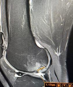
A comprehensive 3T MRI scan of the pelvis and hips provides unparalleled soft-tissue resolution. Unlike standard X-rays which only show bone, a targeted MRI can concurrently evaluate the hip joint for intrinsic causes (like cartilage damage or fluid) while simultaneously visualising the pelvic organs. In cases of occult PID masked as hip pain, the scan may reveal telltale signs of pelvic fluid, tubo-ovarian abscesses, or severe inflammatory stranding that immediately redirects the patient to emergency gynaecological care.
The LSRI Approach: See the Full Picture
Treating presumed tendonitis when a covert pelvic infection is bubbling beneath the surface is the exact scenario our “Diagnose First, Treat Second” philosophy is designed to prevent.
If you have been suffering from unusual, persistent hip or deep pelvic pain that has not responded to conservative therapy, escalating your diagnostic baseline is the safest choice you can make. Do not let referred pain delay critical medical intervention.
The Anatomy Behind Referred Pelvic Pain
The pelvis is one of the most neurologically complex regions of the human body. The obturator nerve, femoral nerve, ilioinguinal nerve, and sacral plexus all converge within a relatively confined anatomical space, creating the conditions for widespread referred pain when any pelvic structure becomes inflamed. Understanding these neural pathways is essential for accurate diagnosis — and is precisely why pelvic MRI is indispensable when hip pain lacks a clear orthopaedic cause.
When to Suspect Non-Orthopaedic Causes of Hip Pain
Certain clinical features should prompt consideration of gynaecological or other non-orthopaedic causes of apparent hip pain:
- Pain that is disproportionate to imaging findings in the hip joint itself
- Cyclical variation in pain intensity correlating with menstrual cycle
- Associated dyspareunia, dysmenorrhoea, or abnormal uterine bleeding
- Absence of localised hip joint tenderness or restricted range of motion
- Previous pelvic inflammatory or gynaecological history
- Bilateral or alternating buttock or groin pain without lumbar pathology
The Role of Advanced Pelvic MRI
Where standard orthopaedic hip imaging (plain X-ray, hip-focused MRI) returns normal or equivocal findings, a comprehensive pelvic MRI provides the definitive next step. This allows simultaneous evaluation of the hip joint, sacroiliac joints, pelvic floor musculature, ovaries, uterus, and bowel — providing a holistic view of the entire anatomical region. Our 3T MRI service at LSRI produces high-resolution pelvic sequences with expert musculoskeletal radiology interpretation, and appropriate onward referral pathways.
A Multidisciplinary Approach to Complex Pelvic Pain
At LSRI, complex cases involving possible non-orthopaedic referred pain are managed with a multidisciplinary lens. Our imaging findings are reported in clinical context, with clear recommendations for onward referral to gynaecology, urology, or gastroenterology where appropriate — ensuring patients do not fall between speciality silos. Book a consultation to discuss your case with our team.

About the Author: Dr Prashant Sankaye, Consultant Musculoskeletal specialist and Radiologist, MBBS, MS, FCPS, MRCS, CCBST, FRCR, PGCE(Med), FHEA, PGDip Sports and Exercise Medicine
Dr Prashant Sankaye is a highly respected Consultant MSK Radiologist and the Clinical Director of London Sports & Rheumatology Imaging (LSRI). With over a decade of sub-specialty experience, he is a recognized expert in advanced diagnostic imaging (Ultrasound & 3T MRI) and precision ultrasound-guided therapeutic injections. His authoritative approach ensures patients avoid surgery where possible and receive the highest standard of orthopaedic, rheumatological, and sports medicine care.