

If you have been referred for an MRI scan — or are considering one — you likely have questions. What exactly is an MRI? How does it differ from a CT scan or X-ray? Should you have a 1.5T or 3T MRI? What if you are claustrophobic?
As a Consultant Radiologist at the London Sports and Rheumatology Imaging (LSRI), Dr Prashant Sankaye sees these questions daily. This comprehensive guide has been written to give patients in London and across the UK a clear, honest, and expert-led understanding of modern MRI scanning — so you can make informed decisions about your health.
What Is an MRI Scan?
Magnetic Resonance Imaging (MRI) is one of the most powerful diagnostic tools in modern medicine. First introduced clinically in the 1980s, MRI uses a combination of strong magnetic fields, radiofrequency pulses, and advanced computer processing to generate highly detailed images of the body's internal structures — without using any ionising radiation.
Unlike X-rays or CT scans, which use radiation to produce images of dense structures like bones, MRI excels at imaging soft tissues — including the brain, spinal cord, muscles, ligaments, tendons, intervertebral discs, and internal organs such as the liver, kidneys, and prostate.
When you have an MRI, you lie inside a large cylindrical magnet. The magnetic field temporarily realigns hydrogen atoms in your body. Radiofrequency pulses are then directed at specific areas, and as those atoms return to alignment, they emit signals that are captured by the scanner and converted into extraordinarily detailed cross-sectional images. The procedure is completely painless and carries no known harmful effects from the magnetic field itself.
Expert Note from Dr Prashant Sankaye: "MRI is arguably the most versatile imaging tool we have. For spinal conditions, neurological disorders, soft tissue injuries, and complex musculoskeletal problems, there is simply no substitute for a high-quality MRI. At LSRI, we ensure every scan is performed to the highest diagnostic standard and reported with precision."
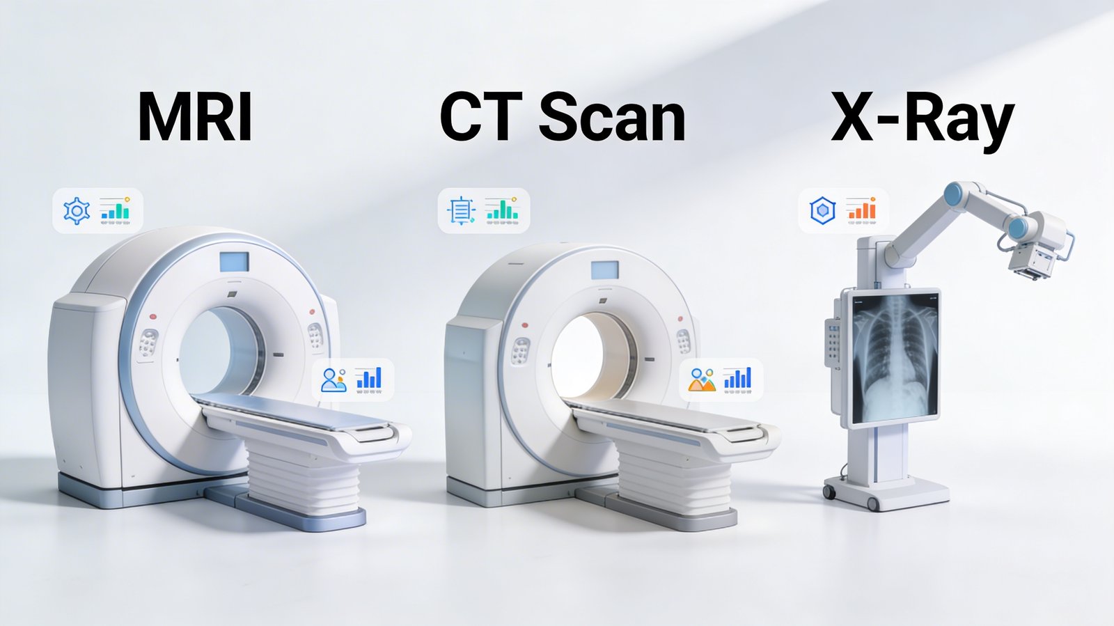
MRI vs CT Scan vs X-Ray: A Full Comparison
Understanding the differences between imaging modalities helps you understand why your doctor chose a specific scan — and empowers you to have better conversations with your medical team.
| Feature | MRI Scan | CT Scan | X-Ray |
|---|---|---|---|
| Technology | Magnetic fields + radio waves | Multiple X-ray beams + computer reconstruction | Single X-ray beam |
| Ionising Radiation | ❌ None | ✅ Yes (moderate-high dose) | ✅ Yes (low dose) |
| Soft Tissue Detail | ⭐⭐⭐⭐⭐ Excellent | ⭐⭐⭐ Good | ⭐ Poor |
| Bone Detail | ⭐⭐⭐ Good | ⭐⭐⭐⭐⭐ Excellent | ⭐⭐⭐⭐ Very Good |
| Brain & Spinal Cord | ⭐⭐⭐⭐⭐ Gold standard | ⭐⭐⭐ Adequate | ❌ Not suitable |
| Ligaments & Tendons | ⭐⭐⭐⭐⭐ Excellent | ⭐⭐ Limited | ❌ Not suitable |
| Lung Detail | ⭐⭐ Limited | ⭐⭐⭐⭐⭐ Excellent | ⭐⭐⭐ Good |
| Abdominal Organs | ⭐⭐⭐⭐⭐ Excellent | ⭐⭐⭐⭐ Very Good | ⭐ Poor |
| Scan Duration | 20–60 minutes | 5–15 minutes | 5 minutes |
| Noise Level | Loud (knocking sounds) | Quiet | Silent |
| Claustrophobia Risk | Moderate-High | Low-Moderate | None |
| Cost (NHS/Private) | Higher | Moderate | Low |
| Best For | Brain, spine, joints, soft tissue | Trauma, chest, complex fractures, abdominal emergencies | Bones, fractures, chest screening |
| Safe in Pregnancy | Second/third trimester with caution | Avoid unless essential | Avoid unless essential |
| Contrast Dye Used | Sometimes (Gadolinium) | Sometimes (Iodine-based) | Rarely |
Key Clinical Takeaways
- MRI is the gold standard for neurological, spinal, musculoskeletal, and pelvic conditions.
- CT is faster and better for trauma, suspected bleeds, or complex bone fractures.
- X-ray remains essential for initial fracture assessment, chest screening, and quick triage.
None of these modalities is "better" universally — each has its clinical purpose. Your radiologist will always recommend the most appropriate scan for your specific situation.
📌 At LSRI, Dr Prashant Sankaye and the clinical team work closely with referring physicians to ensure every patient receives the most diagnostically appropriate and safest imaging pathway.
Related Safety Guide: Considering regenerative medicine? Read our Stem Cell Therapy Patient Safety Guide by Dr Prashant Sankaye.
Types of MRI Scanners: Closed, Wide Bore, Open, 1.5T & 3T
Not all MRI scanners are alike. The type of scanner used can affect image quality, scan time, patient comfort, and clinical outcome. Understanding the options available will help you have an informed discussion with your radiologist.
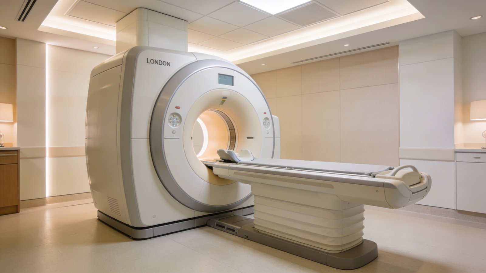
1. Standard Closed Bore MRI
The traditional MRI scanner features a narrow cylindrical tunnel (typically 60–65 cm in diameter) and is what most people picture when they think of an MRI machine.
- Advantages: Highest magnetic field uniformity, reliable image quality, widely available.
- Disadvantages: Can be intimidating for claustrophobic patients, not suitable for larger body types, loud.
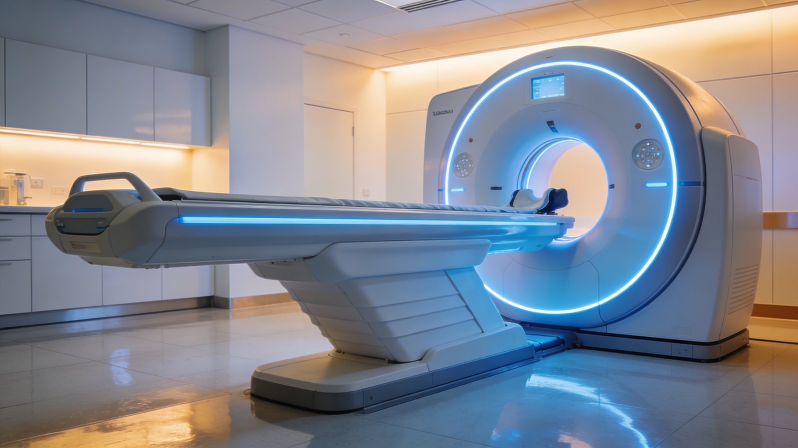
2. Wide Bore MRI Scanner
Wide bore MRI scanners offer a larger tunnel opening — typically 70 cm in diameter — while maintaining the same high diagnostic quality as standard closed bore systems.
- Advantages: Significantly more comfortable, suitable for larger patients, full diagnostic image quality preserved, reduces anxiety.
- Disadvantages: Still enclosed, fewer centres offer wide bore.
Dr Prashant Sankaye's Clinical View: "Wide bore MRI is our preferred scanner for the majority of patients at LSRI. It delivers the diagnostic precision we require while significantly improving patient experience. We rarely compromise on image quality — and with wide bore systems, we don't need to."
3. Open MRI Scanner
Open MRI scanners have no enclosing tunnel at all — the magnet is positioned above and below the patient on an open frame structure, leaving the sides completely open.
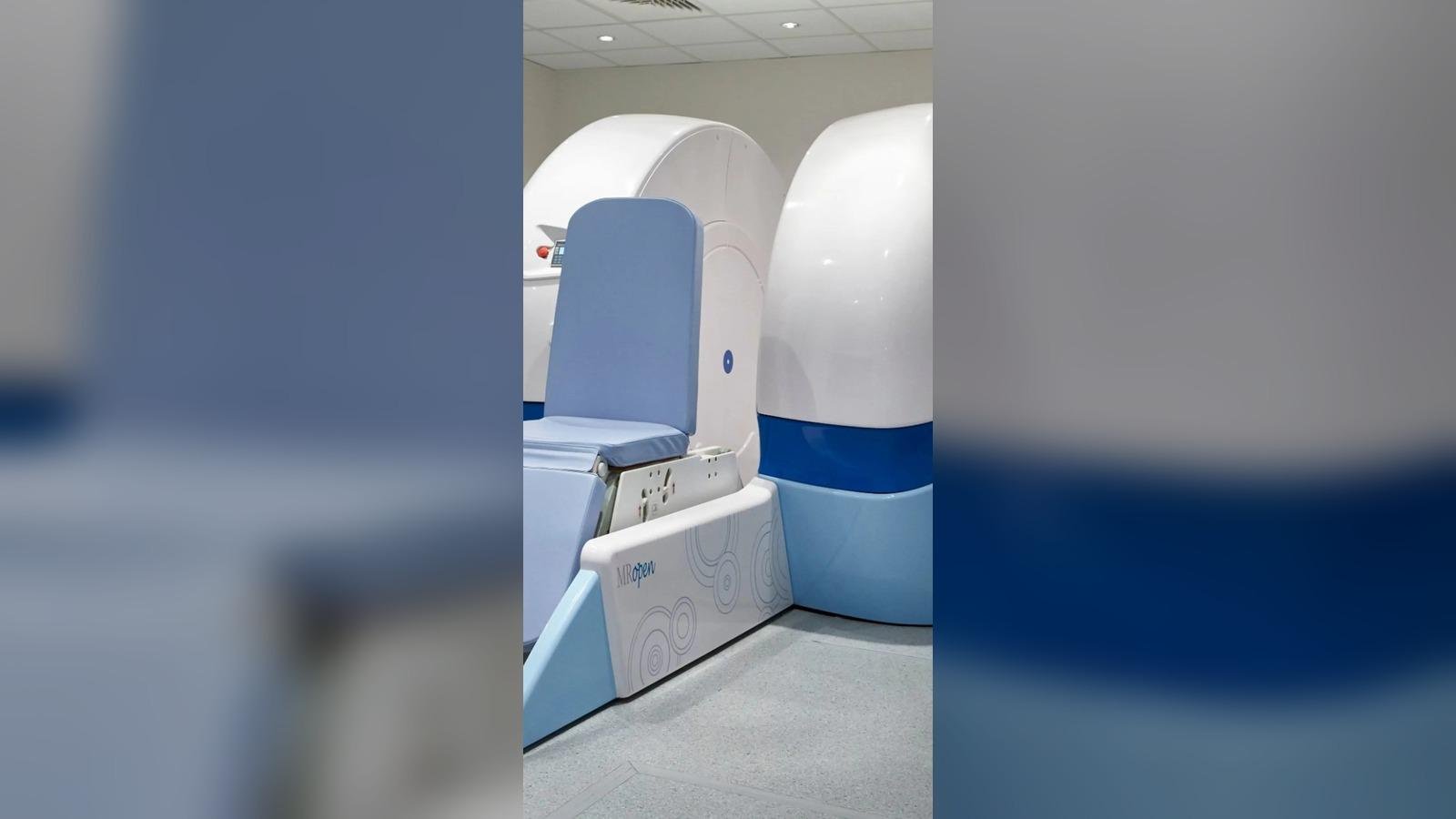
- Advantages: Eliminates claustrophobia entirely, suitable for children who need a parent present.
- Disadvantages: Lower magnetic field strength (0.3T–1.0T), lower image resolution, longer scan times.
⚠️ Important: Open MRI should be considered when patient anxiety or claustrophobia would otherwise prevent a necessary scan. However, if diagnostic image quality is critical, a wide bore closed MRI with appropriate patient preparation is almost always clinically preferable.
4. 1.5 Tesla (1.5T) MRI
Tesla (T) refers to the strength of the magnetic field. A 1.5T MRI has been the clinical workhorse for over three decades.
- Advantages: Proven protocols, excellent general image quality, fewer artefacts from metallic implants.
- Disadvantages: Lower resolution than 3T for fine anatomical detail, longer scan times for equivalent resolution.
5. 3 Tesla (3T) MRI
3T MRI systems offer double the magnetic field strength of 1.5T, translating to significantly higher image resolution, greater signal-to-noise ratio, and faster scan acquisition.
- Advantages: Twice the signal-to-noise ratio, detects subtle pathology, enables advanced techniques (fMRI), exceptional detail for brain and spine.
- Disadvantages: Greater sensitivity to metallic artefacts, more stringent safety screening required.
Which MRI Scanner Is Right for You?
It depends on your clinical condition, your anatomy, your anxiety levels, and what the scan is designed to find. Here is a practical guide:
Choose 3T MRI if:
- Neurological condition investigation
- Complex spinal issues requiring fine detail
- Small joint imaging (wrist, ankle)
- Previous inconclusive 1.5T scan
Choose Wide Bore if:
- Mild claustrophobia
- Larger body type
- You want comfort without sacrificing diagnostic quality
Choose Open MRI if:
- Severe claustrophobia preventing closed scan
- Diagnosed severe anxiety disorders
- Ready to accept lower resolution for comfort
Choose 1.5T if:
- Metallic implants that are less compatible with 3T
- General clinical purpose
- 3T is not clinically indicated for your condition
Always discuss with your radiologist. At LSRI, Dr Prashant Sankaye personally reviews each referral and ensures the most appropriate scanner and protocol is recommended.
MRI Safety: What You Need to Know
MRI is extremely safe, but there are important contraindications that must be screened before every scan.
- Some cardiac pacemakers
- Certain cochlear implants
- Metallic foreign bodies in the eyes
- Some neurostimulators
- Joint replacements/implants (verification needed)
- Claustrophobia (manageable)
- Pregnancy (first trimester avoided)
- Gadolinium contrast allergy
Why Choose LSRI for Your MRI Scan in London?
- ✅ Advanced imaging technology including wide bore and 3T MRI systems.
- ✅ Expert radiologist-led care — Dr Prashant Sankaye and a specialist team.
- ✅ Same-day and next-day appointments available.
- ✅ Rapid, detailed reports — not automated AI summaries.
- ✅ Direct radiologist consultation — speak directly about your results.
- ✅ Transparent pricing — no hidden costs.
Book Your MRI Scan at LSRI London
For complex or urgent cases, patients can request a direct clinical consultation with Dr Prashant Sankaye to discuss the most appropriate imaging pathway.