✍️ Written by: LSRI Editorial Team
🩺 Medically Reviewed by: Dr Prashant Sankaye, Consultant Musculoskeletal specialist and Radiologist, MBBS, MS, FCPS, MRCS, CCBST, FRCR, PGCE(Med), FHEA, PGDip Sports and Exercise Medicine
📅 Last Updated: March 30, 2026
⏱️ Read Time: 4 Minutes
When it comes to Osteochondritis Dissecans Knee, an accurate diagnosis is the first and most critical step toward effective treatment. At LSRI London, we specialize in high-resolution imaging to ensure you receive the precise care your joints need.
When an athlete or active individual sustains a knee injury, the initial response is typically conservative: rest, ice, and physiotherapy. However, when symptoms like persistent pain, swelling, and joint locking persist for months without improvement, an urgent diagnostic review is required. Delaying advanced imaging in these scenarios is not just frustrating for the patient—it can irreversibly alter their long-term prognosis.
A Common Story with Uncommon Consequences
In this anonymised clinical case study, we review a patient who presented to London Sports and Rheumatology Imaging (LSRI) four months after a primary knee injury. Despite undergoing extensive rounds of standard physiotherapy, their mechanical symptoms failed to resolve. They experienced deep-seated knee pain, recurring effusions (swelling), and intermittent locking that severely limited their mobility.
The patient was repeatedly advised to “give it more time” and continue physical therapy. Unfortunately, without a clear view inside the joint, the therapeutic exercises were treating a symptom, not the root pathology.
The 3T MRI Revelation: Osteochondritis Dissecans (OCD)
Upon consultation, Dr Prashant Sankaye immediately recommended a high-resolution 3T MRI scan of the affected knee to definitively map the internal structures. The scan results were striking and sobering.

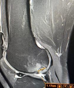
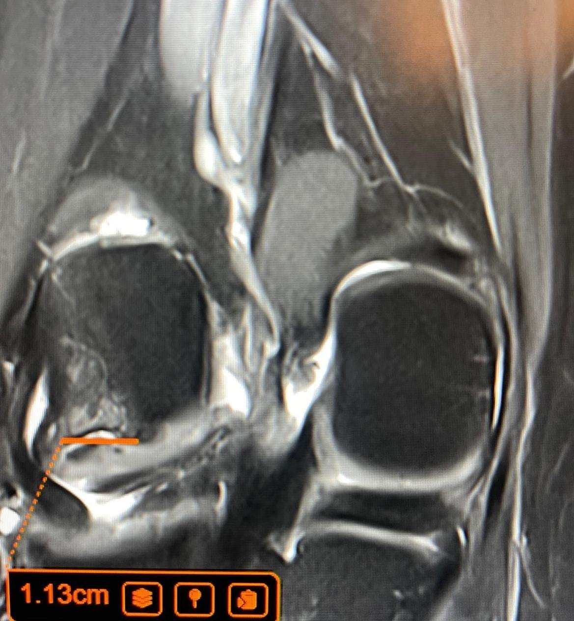
High-resolution MRI scan revealing a severe Osteochondritis Dissecans (OCD) lesion with underlying osteoarthritic changes resulting from prolonged delayed diagnosis.
The imaging revealed an advanced case of Osteochondritis Dissecans (OCD)—a joint condition in which a segment of bone and cartilage loses its blood supply, gradually separating from the underlying bone. Because the joint had been actively loaded and stressed through months of blind rehabilitation efforts, the lesion had deteriorated significantly.
The Price of Delay: Surgery and Osteoarthritis
Had this patient received an MRI scan in the first few weeks following the injury, the OCD lesion could likely have been managed conservatively with strict non-weight-bearing protocols and immobilisation, allowing the bone to heal naturally.
However, due to the 4-month diagnostic delay, the structural damage had progressed past the point of conservative recovery. The MRI scan confirmed that the osteochondral fragment was highly unstable. As a direct result, the patient now requires invasive surgical intervention to secure or remove the loose fragment and stimulate bone healing.
Even more devastatingly, the chronic mechanical stress placed on the compromised joint surface during those four months has triggered the early onset of osteoarthritis, creating a lifelong condition that could have been mitigated.
The Core Lesson: Diagnose First, Treat Second
This case fiercely underscores Dr Sankaye’s clinical philosophy: Diagnose first, treat second.
Mechanical symptoms such as joint locking, giving way, or persistent swelling that does not respond to initial basic care are massive red flags. Pushing through the pain or blindly continuing physiotherapy without a definitive anatomical map is clinically dangerous. Early imaging eliminates the guesswork, ensures therapies are safely targeted, and—most importantly—prevents treatable injuries from evolving into permanent, life-altering degenerative conditions.
If you or a patient have been struggling with an unresolved MSK injury for longer than expected, do not wait until structural failure occurs. Secure an expert diagnostic ultrasound or MRI scan to achieve complete clinical clarity.
Long-Term Care and Prognosis
Early diagnosis is the most critical factor in achieving excellent clinical outcomes. Advanced imaging prevents minor degenerative changes from progressing into chronic, debilitating conditions. Patients undergoing specialist musculoskeletal imaging immediately receive diagnostic clarity, followed by appropriate, targeted interventions such as ultrasound-guided injections or sophisticated rehabilitative plans. Dr Prashant Sankaye and the LSRI experts emphasise precise anatomical correlation to ensure symptoms are accurately treated rather than temporarily masked.
About the Author: Dr Prashant Sankaye, Consultant Musculoskeletal specialist and Radiologist, MBBS, MS, FCPS, MRCS, CCBST, FRCR, PGCE(Med), FHEA, PGDip Sports and Exercise Medicine
Dr Prashant Sankaye is a highly respected Consultant MSK Radiologist and the Clinical Director of London Sports & Rheumatology Imaging (LSRI). With over a decade of sub-specialty experience, he is a recognized expert in advanced diagnostic imaging (Ultrasound & 3T MRI) and precision ultrasound-guided therapeutic injections. His authoritative approach ensures patients avoid surgery where possible and receive the highest standard of orthopaedic, rheumatological, and sports medicine care.