Written by: LSRI Editorial Team
Medically Reviewed by: Dr Prashant Sankaye, Consultant MSK Radiologist — MBBS, MS, FCPS, MRCS, CCBST, FRCR, PGCE(Med), FHEA, PGDip Sports and Exercise Medicine
Last Updated: October 14, 2026
Read Time: 4 Minutes
A shoulder MRI scan provides detailed soft tissue imaging that no other scan can match for depth and comprehensiveness. If you have persistent shoulder pain, a clicking or catching sensation in your shoulder, weakness when lifting, or pain that is not improving with physiotherapy or anti-inflammatories, a shoulder MRI scan is likely the most important investigation you can have. This guide explains exactly what a shoulder MRI detects, how it compares to ultrasound and X-ray, what happens during the scan, and when the findings directly change your treatment plan.
📋 In This Article
What Can a Shoulder MRI Scan Detect?
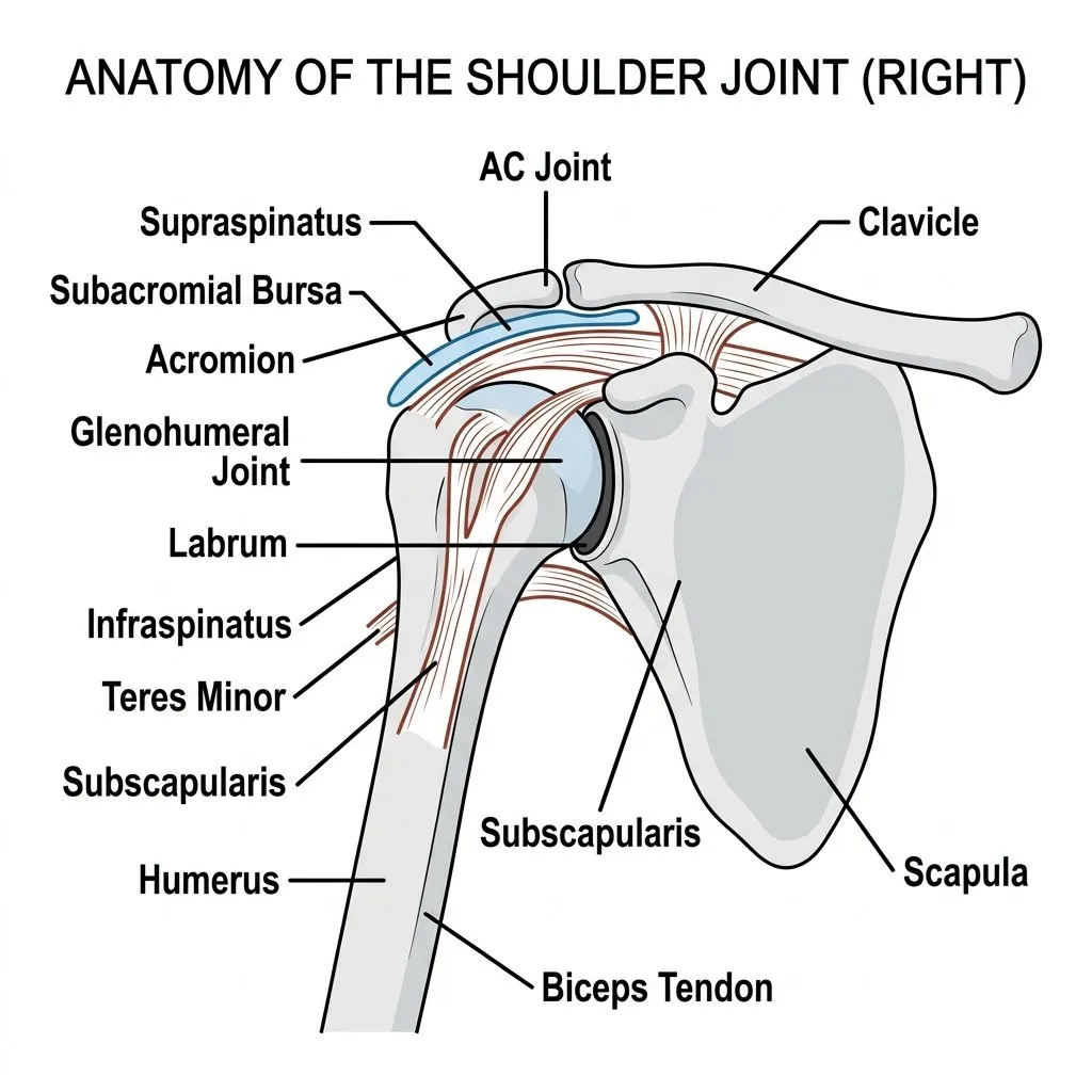
An MRI (Magnetic Resonance Imaging) scan uses powerful magnetic fields and radio waves to create highly detailed cross-sectional images of the shoulder. Unlike X-rays, which only show bone, MRI shows the muscles, tendons, ligaments, cartilage, labrum, and bone marrow in exquisite detail.
At London Sports and Rheumatology Imaging, our MRI protocols are specifically optimised for musculoskeletal diagnosis. A shoulder MRI can definitively identify:
- Rotator Cuff Tears: Whether partial or full-thickness, MRI shows the exact size of the tear, the degree of tendon retraction, and — crucially for surgeons — whether the associated muscle has begun to atrophy (waste away).
- Labral Tears: The labrum is the ring of cartilage surrounding the shoulder socket. SLAP tears and Bankart lesions (often caused by dislocation) are visualised clearly.
- Capsular Problems: The thickening and contraction of the joint capsule characteristic of frozen shoulder (adhesive capsulitis).
- Bone and Cartilage Damage: Osteoarthritis, bone bruising (often seen after injury), and avascular necrosis.
- Bursal and Tendon Inflammation: Subacromial bursitis and biceps tendinopathy.
MRI vs Ultrasound vs X-Ray: Which Do You Need?
These three investigations are complementary, not competing. At LSRI, we often combine ultrasound for dynamic real-time assessment with MRI for deep structure evaluation.
- X-ray: Best for fractures, severe osteoarthritis, and detecting calcific tendinopathy. It does not show tendons or cartilage.
- Ultrasound: Best for real-time, dynamic assessment of the rotator cuff and bursa (watching them move), and for guiding injections. It is fast and highly accurate for superficial structures but cannot see deep into the joint (e.g., the labrum).
- MRI: The gold standard for seeing the entire shoulder joint, deep cartilage, the labrum, and bone marrow. It is a static image but provides unparalleled detail.
When Should You Have a Shoulder MRI?
You should consider a shoulder MRI if you have:
- Shoulder pain lasting more than 6 weeks
- Suspected rotator cuff tear (weakness when lifting the arm)
- A history of shoulder dislocation or instability
- Severe night pain
- Pain following a specific injury or trauma
- Symptoms that have not resolved with physiotherapy or previous steroid injections
- A surgical referral pending — surgeons require an MRI before planning an operation
What Happens During a Shoulder MRI at LSRI?
- The scan typically takes 20-30 minutes.
- You will lie on a scanning bed, and a specific “coil” (a specialized receiver) is placed over your shoulder.
- The scanner makes loud tapping noises; you will be provided with ear protection and can often listen to music.
- The scan is completely painless and involves no radiation.
Following your scan, Dr Prashant Sankaye will analyse the images and provide a comprehensive specialist report. Depending on the findings, we can immediately discuss non-surgical treatment options such as hydrodistension, ultrasound-guided steroid injections, or PRP.
A private shoulder MRI at LSRI costs from £375, including a report from a consultant MSK radiologist within 24 hours. More about our private shoulder MRI.
For more information, see our pages on rotator cuff tears and frozen shoulder.
Dr Prashant Sankaye is a specialist musculoskeletal radiologist and CPD Approved Trainer at London Sports and Rheumatology Imaging (LSRI Ltd), CPD Provider #790052.