Written by: LSRI Editorial Team
Medically reviewed by Dr Prashant Sankaye, Consultant MSK Radiologist ·
Last Updated: October 14, 2026
Read Time: 17 Minutes
Written by the LSRI clinical team and medically reviewed by Dr Prashant Sankaye, Consultant MSK Radiologist, on 10 October 2026.
Many twisted knees settle with time and simple care. A few do not, and they often show it early: the knee locks and will not straighten, it gives way when you turn, or it swells up within hours. These signs point to damage inside the joint, such as a torn meniscus or a torn anterior cruciate ligament (ACL). The menisci are two rubbery, C-shaped cushions between the thigh bone and shin bone. The ACL, in the centre of the knee, stops the shin bone sliding forwards and twisting.
This article explains how to tell a truly locked knee from a stiff one, which warning signs need a prompt check, and when an X-ray or MRI helps. The aim is to know the diagnosis early, so that treatment is chosen on purpose. Our patient guide to knee MRI explains what the scan involves.
Key points
- A truly locked knee cannot be fully straightened because something inside the joint is in the way, most often a displaced “bucket-handle” meniscal tear; pain and muscle spasm can mimic this1.
- UK knee surgeons recommend urgent arthroscopic (keyhole) surgery for a locked knee2.
- A knee that gives way or becomes badly swollen after an injury should be checked promptly3. In one UK study, only 28% of ACL tears were correctly diagnosed at the first medical visit4.
- The Ottawa knee rules show which adults need an X-ray; in a systematic review, the rule picked up 98.5% of fractures5,6.
- Long delays before ACL surgery are linked with more meniscal damage7, yet rehabilitation first, with surgery only if needed, gave similar five-year results in a randomised trial8.
- Meniscal damage is common on MRI in adults over 50, and in one community study most of it had caused no recent pain9, so a scan needs to be read alongside your symptoms and examination.
What does a truly locked knee feel like?
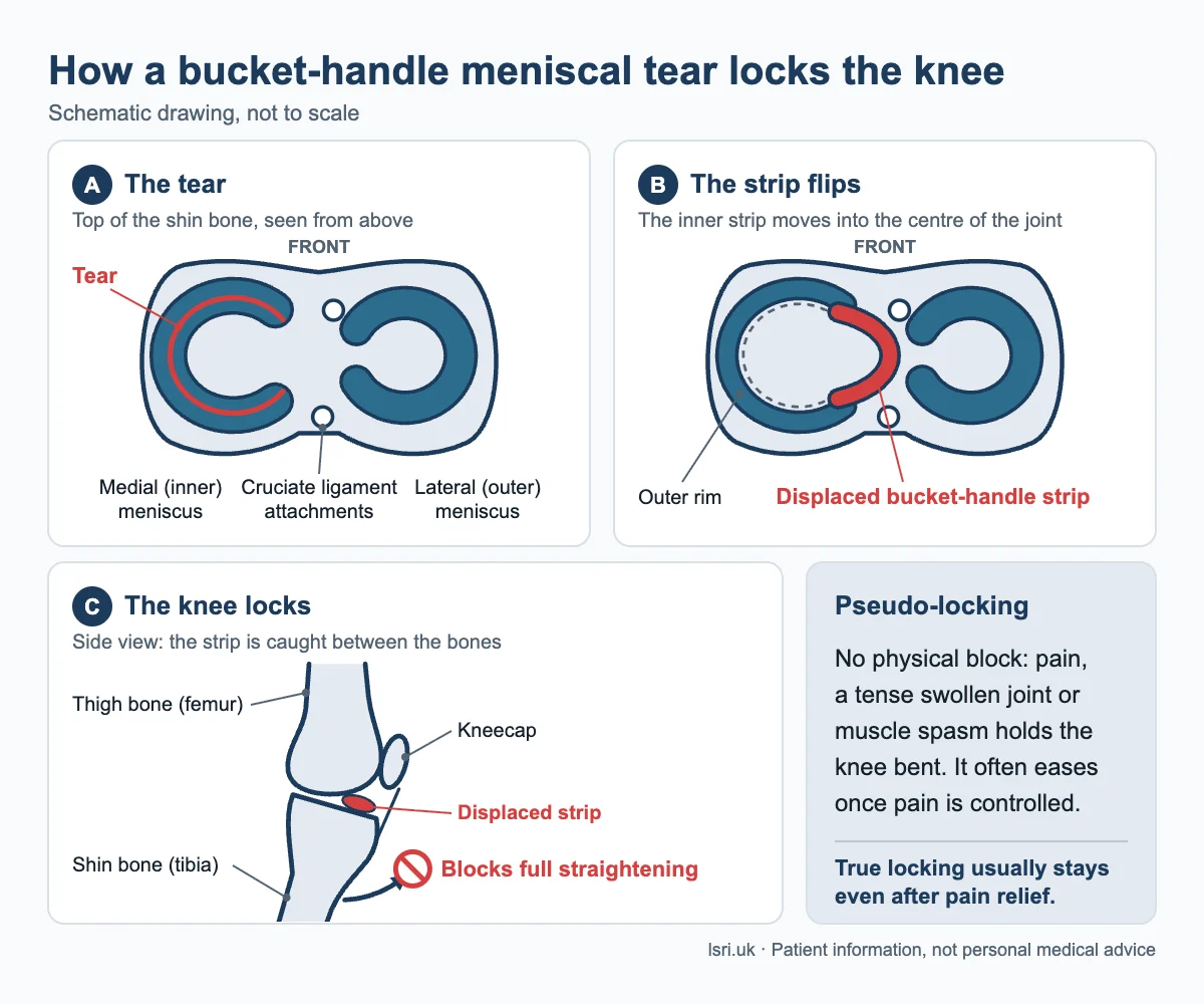
A truly locked knee is stuck short of straight. It may still move a little, but something inside the joint blocks full straightening, and the block usually remains even after pain relief1.
The most common physical cause is a torn meniscus, classically a bucket-handle tear1. This is a long split along the meniscus whose inner strip flips into the middle of the joint, like the handle of a bucket lifting up, and jams between the bones. Other causes include a loose piece of bone or cartilage, for example from osteochondritis dissecans, and a torn ACL1.
Not every knee that will not straighten is truly locked. In pseudo-locking, pain, a tense swollen joint or muscle spasm holds the knee bent without any physical block. An older estimate quoted in a recent review puts this at up to half of knees that arrive as “locked”; it often eases once pain is controlled1.
Clicking and catching are different again. Painless clicking is normal3. In people with a degenerative (wear-related) meniscal tear, keyhole surgery relieved catching or occasional locking no better than a placebo (sham) operation10. That finding does not apply to a knee that is fixed and cannot straighten10.
| True locking | Pseudo-locking | Catching or clicking | |
|---|---|---|---|
| What you notice | Stuck short of straight | Held bent by pain, swelling or spasm | A brief catch or click, then normal movement |
| Typical cause | Displaced meniscal tear, loose fragment or torn ACL1 | Pain and muscle guarding, no physical block1 | In wear-related tears, surgery on the tear helped no more than sham surgery10 |
| What it needs | Prompt specialist assessment, usually with MRI2 | Assessment; often eases once pain is controlled1 | Advice if painful, or with locking, giving way or bad swelling3 |
What does it mean if your knee gives way?
Giving way means the knee suddenly buckles, often when you turn or land. After a twisting injury, a torn ACL is a common cause, and so is a dislocated kneecap (patellar dislocation), where the kneecap slips out of its groove, usually to the outer side. Both were common injuries in a large MRI study of 1,145 acutely injured knees11.
An ACL tear can be missed. In a UK study of 117 ACL tears treated with surgery (median age 18), three in four followed a typical injury pattern, yet only 28% were correctly diagnosed at the first medical visit4. One reason is that bedside tests are less reliable in an awake patient. In a review of 20 studies of acute complete ACL tears, the pivot shift test (a twisting test) picked up 28% of tears in awake patients and 73% under anaesthesia, and the Lachman test (a forward pull on the shin) 81% overall and 91% under anaesthesia12.
Do you need an X-ray first? The Ottawa knee rules
An X-ray looks for broken bones. After an acute knee injury in an adult, the Ottawa knee rules advise an X-ray if any one of these applies5:
- age 55 or over
- tenderness at the head of the fibula (the bony bump on the outer side, just below the knee)
- tenderness of the kneecap alone, with no other bony tenderness
- you cannot bend the knee to 90 degrees
- you cannot take four steps (two on each leg, limping allowed), both straight after the injury and when examined
A systematic review of six studies (4,249 adults) found that the rule picked up 98.5% of fractures, so a negative result makes a fracture very unlikely6. It is deliberately cautious: about half of people without a fracture still meet a criterion6. It was designed for adults; its use in children is less well established5.
An X-ray cannot show the menisci, ligaments or cartilage, and it may be normal after a purely soft-tissue injury, so it does not rule out a torn ACL or meniscus1.
When does an MRI add real value?
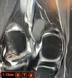
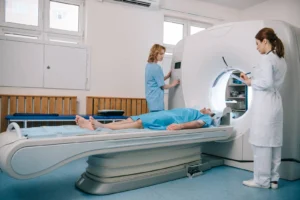
MRI (magnetic resonance imaging) uses a strong magnet and radio waves, not X-rays, to show the menisci, ligaments, cartilage and bone. A European expert consensus calls it a useful, highly accurate test before surgery. The experts did not agree that every traumatic meniscal tear needs one, and advised that a musculoskeletal specialist should decide when it is needed13. It adds most when the answer will change what happens next:
- A knee that will not straighten. UK guidance notes that most people with a locked knee will have a displaced meniscal tear confirmed on MRI2.
- A suspected ACL tear, for example a twist followed by rapid swelling or giving way. Bedside tests can miss ACL tears12, and most ACL tears came with other injuries11.
- Catching or giving way that persists after an injury, or a diagnosis that is still unclear.
- Planning surgery, so the surgeon knows whether a meniscus may be repairable and what else is injured13.
MRI is accurate but not perfect. In a meta-analysis of studies comparing MRI with arthroscopy (keyhole surgery), MRI detected 87% of ACL tears, 89% of medial (inner) meniscal tears and 78% of lateral (outer) meniscal tears, and correctly cleared 88% to 95% of knees without a tear14. Most of the studies had a high or unclear risk of bias14, and these figures are not a head-to-head comparison with the bedside tests above. That is why a scan should be read alongside the history and examination. Ultrasound is not the usual test for these injuries: in the same review, there were too few ultrasound studies to pool14.
What can a delayed diagnosis cost?
The main worry with a missed ACL or meniscal injury is not missing a window for surgery. It is possible further damage while the knee keeps giving way, and a lower chance of repairing a meniscus rather than removing part of it1,7. The studies below show links, not proof of cause. The same principle applies to musculoskeletal injuries more widely.
- In the UK study above, when diagnosis took more than six months, 72% had a medial meniscal tear, compared with 23% when it was made within four months4.
- In a US registry of 1,252 people having ACL reconstruction, surgery at 6 to 12 months carried 1.8 times the odds of a medial meniscus injury compared with earlier surgery, and 2.2 times after 12 months7.
This does not mean everyone needs early surgery. A randomised trial of 121 active adults aged 18 to 35, with an ACL tear no more than four weeks old, gave everyone structured rehabilitation, plus either early reconstruction or later reconstruction only if the knee stayed unstable8. At five years, knee scores were similar, with no significant difference in meniscal surgery or X-ray signs of arthritis, though tests of knee looseness were better after early surgery8. Of the rehabilitation-first group, 49% had not had a reconstruction8. The lesson is not “operate early”. It is “know early”, so that rehabilitation, surgery or both are chosen deliberately.
A locked knee is more pressing. UK guidance recommends urgent surgery2, European guidance advises that a traumatic tear needing repair should be repaired as early as possible13, and a review notes that the chance of a successful repair falls the later a locked knee presents1. The research on exact timing is less clear: in a meta-analysis of 38 studies of bucket-handle repairs, about 15% failed, and time from injury to surgery did not significantly predict failure15. A knee that cannot straighten needs a diagnosis and a plan without delay.
Is an MRI worth it for a minor twist or gradual knee pain?
It can be, but the scan needs to be read alongside your symptoms and examination. This matters most in people over about 50 whose pain came on gradually, because meniscal changes on MRI are common at that age, often without pain.
In a community study of 991 people aged 50 to 90, MRI showed meniscal damage (a tear or a worn-away meniscus) in 19% of women aged 50 to 59, rising to 56% in men aged 70 to 909. Of the knees with meniscal damage, 61% had had no pain, aching or stiffness in the previous month9. A tear on a scan does not prove it is causing the pain.
For wear-related (degenerative) meniscal tears, a Finnish trial of 146 people aged 35 to 65 without arthritis found that keyhole surgery to trim the tear was no better at 12 months than a sham operation16. European and UK guidance advise non-surgical treatment first, for at least three months, before keyhole surgery is considered, with earlier surgery for a locked knee2,17.
Two common situations:
- Gradual pain, without injury, over about 50: a standing X-ray usually comes first17 (see our knee osteoarthritis page). An MRI can help if symptoms do not settle or another problem is suspected.
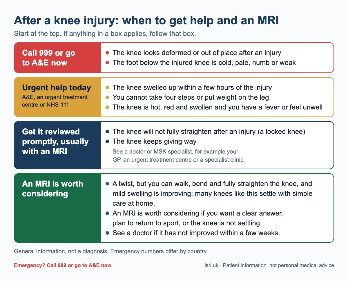
- A twist, but you can walk, bend and fully straighten the knee, and mild swelling is improving: many knees like this settle with simple care at home. In our view, an MRI gives the clearest picture of what has been injured, and is worth considering if you want a clear answer, plan to return to sport, or the knee is not settling. See a doctor if it has not improved within a few weeks3.
What to do after a twisting knee injury
Go to A&E or call 999 now if you have:
- a knee that looks deformed or out of place after an injury
- a foot below the injured knee that is cold, pale, numb or weak
Get urgent medical help today (A&E, an urgent treatment centre or NHS 111) if:
- the knee swelled up within a few hours of the injury
- you cannot take four steps or put weight on the leg
- the knee is hot, red and swollen and you have a fever or feel unwell (read about the hot, swollen joint)
A dislocated knee can damage the blood vessels and nerves around it. In a systematic review of 862 people with knee dislocation, about 18% had a blood vessel injury, and 25% of those assessed had a nerve injury18.
If your knee will not fully straighten after an injury (a locked knee), get it reviewed promptly by a doctor or musculoskeletal (MSK) specialist, for example your GP, an urgent treatment centre or a specialist clinic. It usually needs an MRI to find the cause2.
- Check for red flags first. If anything in the box above applies, get urgent care rather than booking a scan.
- Get an X-ray if the Ottawa knee rules point to one. One criterion is enough5.
- Ask a doctor whether an MRI would help if the knee locks, gives way or swelled quickly. An accurate diagnosis can change the plan for these injuries2,11.
- Do not wait months with a knee that keeps giving way. One UK study recommended that people with a typical ACL injury see a knee specialist within six weeks4.
- Get it checked again if things change. New locking, giving way, marked swelling, or a hot knee with fever need prompt review3.
What does treatment look like once the diagnosis is clear?
Treatment follows the diagnosis; our knee meniscal tear page has more on tears.
- Locked knee from a displaced meniscal tear: urgent keyhole surgery to free the knee2, repairing the meniscus where possible13.
- ACL tear: structured rehabilitation, with reconstruction either early or later if the knee stays unstable8; the choice is made with your surgeon.
- Dislocated kneecap: the plan depends on the scan, including any loose fragment of cartilage or bone, which can cause locking1.
How LSRI can help
LSRI is a doctor-led musculoskeletal (MSK) imaging clinic, not an emergency service. If anything in the red box above applies, get urgent care first.
For a knee injury that is not an emergency, we offer:
- Knee MRI reported by a consultant MSK radiologist within 24 hours. MRI scans start from £375 per body part, including the scan, a specialist report and access to your images (from £425 with a consultation).
- Ultrasound for some soft-tissue questions around the knee, such as tendon problems.
- A second opinion on a knee MRI you have already had: see our second opinion page.
Our clinics are on Harley Street (W1G 9QJ) and in Harrow (HA2 7SJ); call 0203 633 5040.
Reading this from outside the UK?
The medical points here apply everywhere, but emergency numbers differ: 999 or 112 in the UK, 112 across the EU, 911 in the US and Canada, and 000 in Australia. NHS 111 is a UK service; elsewhere, use your local urgent care service.
Frequently asked questions
Can a torn meniscus lock your knee?
Yes. A torn meniscus, especially a bucket-handle tear, is the most common physical cause of a truly locked knee1, and UK guidance recommends urgent keyhole surgery2.
How do I know if my knee is locked or just stiff?
A truly locked knee will not straighten fully, usually even after pain relief, because something inside it is in the way1. In pseudo-locking, pain or spasm holds it bent, and it often eases with pain relief1. If you cannot fully straighten your knee after an injury, get it reviewed promptly by a doctor or MSK specialist; a locked knee usually needs an MRI2.
Can an ACL tear get better without surgery?
Some people do well without reconstruction. In a randomised trial of young, active adults, 49% of those who started with rehabilitation had not needed reconstruction at five years, with knee scores similar to early surgery8. A knee that keeps giving way is a reason to discuss surgery, as long delays are linked with more meniscal damage4,7.
Should I get an MRI for knee pain if I am over 50?
A standing X-ray is usually the first test for knee pain that came on gradually17. An MRI can help if symptoms do not settle, you want a clear answer, or another problem is suspected, but meniscal damage is common on MRI at this age, often without pain9, so the scan needs to be read alongside your symptoms.
References
- Rhind JH, Khawar H, Webb M, Guthrie H. The locked knee. Br J Hosp Med (Lond) 2024;85(1):1-9. doi:10.12968/hmed.2022.0215
- Abram SGF, Beard DJ, Price AJ; BASK Meniscal Working Group. Arthroscopic meniscal surgery: a national society treatment guideline and consensus statement. Bone Joint J 2019;101-B(6):652-659. doi:10.1302/0301-620X.101B6.BJJ-2019-0126.R1
- NHS. Knee pain. nhs.uk (page last reviewed 21 December 2023; accessed 8 October 2026)
- Arastu MH, Grange S, Twyman R. Prevalence and consequences of delayed diagnosis of anterior cruciate ligament ruptures. Knee Surg Sports Traumatol Arthrosc 2015;23(4):1201-1205. doi:10.1007/s00167-014-2947-z
- Yao K, Haque T. The Ottawa knee rules: a useful clinical decision tool. Aust Fam Physician 2012;41(4):223-224. racgp.org.au
- Bachmann LM, Haberzeth S, Steurer J, ter Riet G. The accuracy of the Ottawa knee rule to rule out knee fractures: a systematic review. Ann Intern Med 2004;140(2):121-124. doi:10.7326/0003-4819-140-5-200403020-00013
- Chhadia AM, Inacio MCS, Maletis GB, Csintalan RP, Davis BR, Funahashi TT. Are meniscus and cartilage injuries related to time to anterior cruciate ligament reconstruction? Am J Sports Med 2011;39(9):1894-1899. doi:10.1177/0363546511410380
- Frobell RB, Roos HP, Roos EM, Roemer FW, Ranstam J, Lohmander LS. Treatment for acute anterior cruciate ligament tear: five year outcome of randomised trial. BMJ 2013;346:f232. doi:10.1136/bmj.f232
- Englund M, Guermazi A, Gale D, Hunter DJ, Aliabadi P, Clancy M, et al. Incidental meniscal findings on knee MRI in middle-aged and elderly persons. N Engl J Med 2008;359(11):1108-1115. doi:10.1056/NEJMoa0800777
- Sihvonen R, Englund M, Turkiewicz A, Järvinen TLN; Finnish Degenerative Meniscal Lesion Study Group. Mechanical symptoms and arthroscopic partial meniscectomy in patients with degenerative meniscus tear: a secondary analysis of a randomized trial. Ann Intern Med 2016;164(7):449-455. doi:10.7326/M15-0899
- Olsson O, Isacsson A, Englund M, Frobell RB. Epidemiology of intra- and peri-articular structural injuries in traumatic knee joint hemarthrosis: data from 1145 consecutive knees with subacute MRI. Osteoarthritis Cartilage 2016;24(11):1890-1897. doi:10.1016/j.joca.2016.06.006
- van Eck CF, van den Bekerom MPJ, Fu FH, Poolman RW, Kerkhoffs GMMJ. Methods to diagnose acute anterior cruciate ligament rupture: a meta-analysis of physical examinations with and without anaesthesia. Knee Surg Sports Traumatol Arthrosc 2013;21(8):1895-1903. doi:10.1007/s00167-012-2250-9
- Kopf S, Beaufils P, Hirschmann MT, Rotigliano N, Ollivier M, Pereira H, et al. Management of traumatic meniscus tears: the 2019 ESSKA meniscus consensus. Knee Surg Sports Traumatol Arthrosc 2020;28(4):1177-1194. doi:10.1007/s00167-020-05847-3
- Phelan N, Rowland P, Galvin R, O’Byrne JM. A systematic review and meta-analysis of the diagnostic accuracy of MRI for suspected ACL and meniscal tears of the knee. Knee Surg Sports Traumatol Arthrosc 2016;24(5):1525-1539. doi:10.1007/s00167-015-3861-8
- Costa GG, Grassi A, Zocco G, Graceffa A, Lauria M, Fanzone G, et al. What is the failure rate after arthroscopic repair of bucket-handle meniscal tears? A systematic review and meta-analysis. Am J Sports Med 2022;50(6):1742-1752. doi:10.1177/03635465211015425
- Sihvonen R, Paavola M, Malmivaara A, Itälä A, Joukainen A, Nurmi H, et al. Arthroscopic partial meniscectomy versus sham surgery for a degenerative meniscal tear. N Engl J Med 2013;369(26):2515-2524. doi:10.1056/NEJMoa1305189
- Beaufils P, Becker R, Kopf S, Englund M, Verdonk R, Ollivier M, et al. Surgical management of degenerative meniscus lesions: the 2016 ESSKA meniscus consensus. Knee Surg Sports Traumatol Arthrosc 2017;25(2):335-346. doi:10.1007/s00167-016-4407-4
- Medina O, Arom GA, Yeranosian MG, Petrigliano FA, McAllister DR. Vascular and nerve injury after knee dislocation: a systematic review. Clin Orthop Relat Res 2014;472(9):2621-2629. doi:10.1007/s11999-014-3511-3
Book an appointment
Call 0203 633 5040 or book online. Clinics at Harley Street (W1G 9QJ) and Harrow (HA2 7SJ). If you have emergency symptoms, go to A&E or call 999 instead.
About this article
Purpose: written by the LSRI clinical team to help the public recognise the knee injury warning signs (a locked knee, a knee that gives way and rapid swelling) that need a prompt, accurate diagnosis, and what a knee X-ray or MRI can and cannot show, based on the research and guidance listed above. It is general information, not a diagnosis. If you are worried about your own symptoms, speak to your GP or, in an emergency, go to A&E. This article does not describe any individual patient and is not a criticism of any clinician.