Written by: LSRI Editorial Team
Medically reviewed by Dr Prashant Sankaye, Consultant MSK Radiologist ·
Last Updated: October 14, 2026
Read Time: 17 Minutes
Low back pain is extremely common, and most of it is mechanical: it comes from muscles, joints and discs, and it improves with time. But in some people, particularly older adults, back and buttock pain that will not settle comes from a crack in the sacrum, the triangular bone at the base of the spine. These sacral insufficiency fractures happen in weakened bone under everyday load, often without any fall or injury. They are hard to see on X-ray and easy to mistake for ordinary back pain or sciatica. Just as importantly, they are a warning sign: a bone that breaks this easily usually means fragile bones elsewhere too. This anonymised case from our clinic, in which three months of back pain turned out to be a sacral insufficiency fracture, explains the condition, why MRI matters, and what to do next about bone health, including osteoporosis, DEXA scans, vitamin D and medicines such as alendronic acid.
Key points
- A sacral insufficiency fracture is a crack in the sacrum caused by normal, everyday stress on bone weakened by osteoporosis or other conditions.1
- It usually causes low back, buttock, hip or groin pain, sometimes with leg pain like sciatica, so it is often mistaken for common back problems.1,2
- Plain X-rays detect only about 20–38% of these fractures. MRI detects close to all of them.1
- Most heal with pain relief, careful mobilisation and time.1 Manipulation of the spine and pelvis needs caution in osteoporosis and is not used on an acute fracture.3
- Any fragility fracture should lead to a bone health check: risk assessment, usually a DEXA scan, and, where needed, treatment to prevent the next fracture.4
The case: three months of back and buttock pain
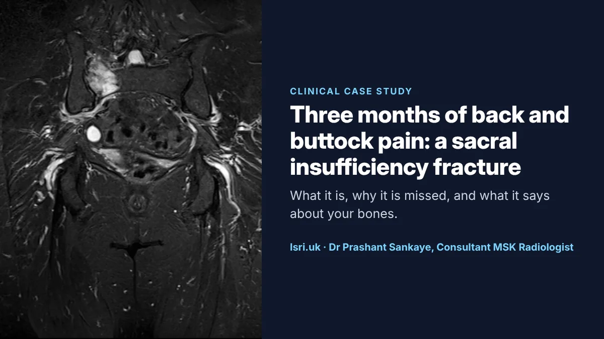
A patient developed pain in the lower back and buttock. Over about three months it was treated as mechanical back pain, without lasting improvement. An MRI scan at LSRI showed a sacral insufficiency fracture on the right side of the sacrum, with swelling in the surrounding bone marrow.
This case is not a criticism of the practitioners involved. Without a scan, an insufficiency fracture of the sacrum is very hard to tell apart from common back pain: the symptoms overlap, there is often no injury to explain them, and a plain X-ray is usually normal. The lesson is about the pathway: when back pain in someone at risk of fragile bones does not follow the expected course, the diagnosis needs to be looked at again.
What the MRI showed
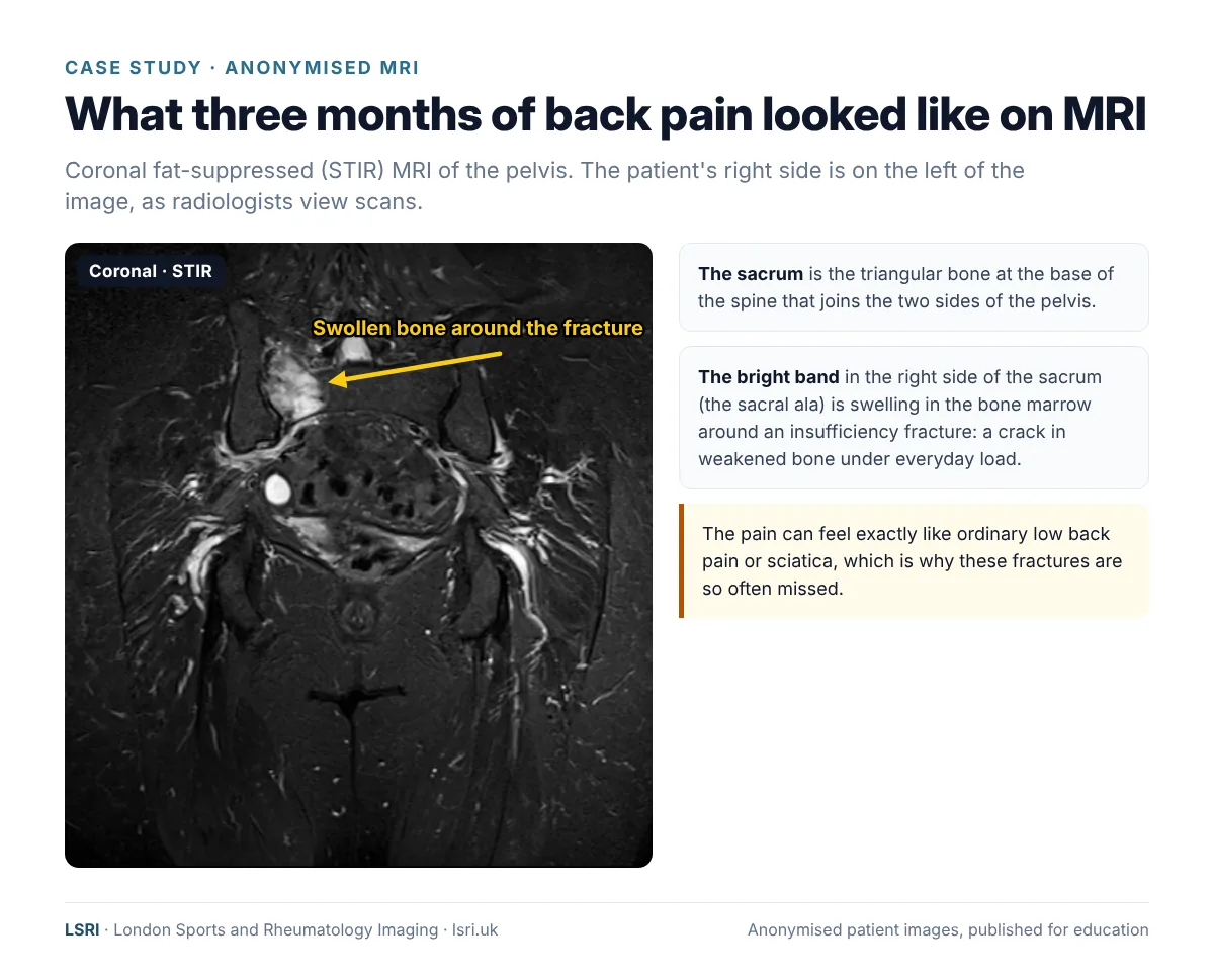
This image uses a STIR sequence, which darkens fat so that water stands out. Healthy adult bone marrow contains a lot of fat and looks dark on STIR. Around a fracture, the marrow fills with fluid (oedema) and lights up. Here, a bright band runs through the right side of the sacrum, the part called the sacral ala, where insufficiency fractures typically occur. In about half of cases both sides of the sacrum are affected, sometimes with a horizontal fracture joining them.1,5
What is a sacral insufficiency fracture?
There are two broad kinds of stress fracture. Fatigue fractures happen when normal bone is overloaded, for example in runners. Insufficiency fractures happen when bone is weakened, so that the ordinary forces of standing, walking and getting up from a chair are enough to crack it.1,6 The sacrum carries the weight of the upper body to the pelvis and legs, which makes it a common site.
Who is at risk?
- Osteoporosis: the most common underlying cause, particularly in women after the menopause.1
- Long-term steroid treatment, and conditions such as rheumatoid arthritis.1
- Radiotherapy to the pelvis: in a review of nearly 4,000 women treated with pelvic radiotherapy for gynaecological cancers, about 14% developed pelvic insufficiency fractures.7
- A previous fragility fracture, such as a broken wrist, spine or hip after a minor fall.
Symptoms
The pain is usually in the lower back, buttock, hip or groin, and it is typically worse with standing and walking and better lying down. Some people have pain spreading into the leg, which can look exactly like sciatica from a disc problem. There is often no fall or injury, or only a very minor one.1,2 There is often a delay of around six to eight weeks from the start of symptoms before the sacrum is specifically imaged.1
Why X-rays miss it, and why MRI finds it
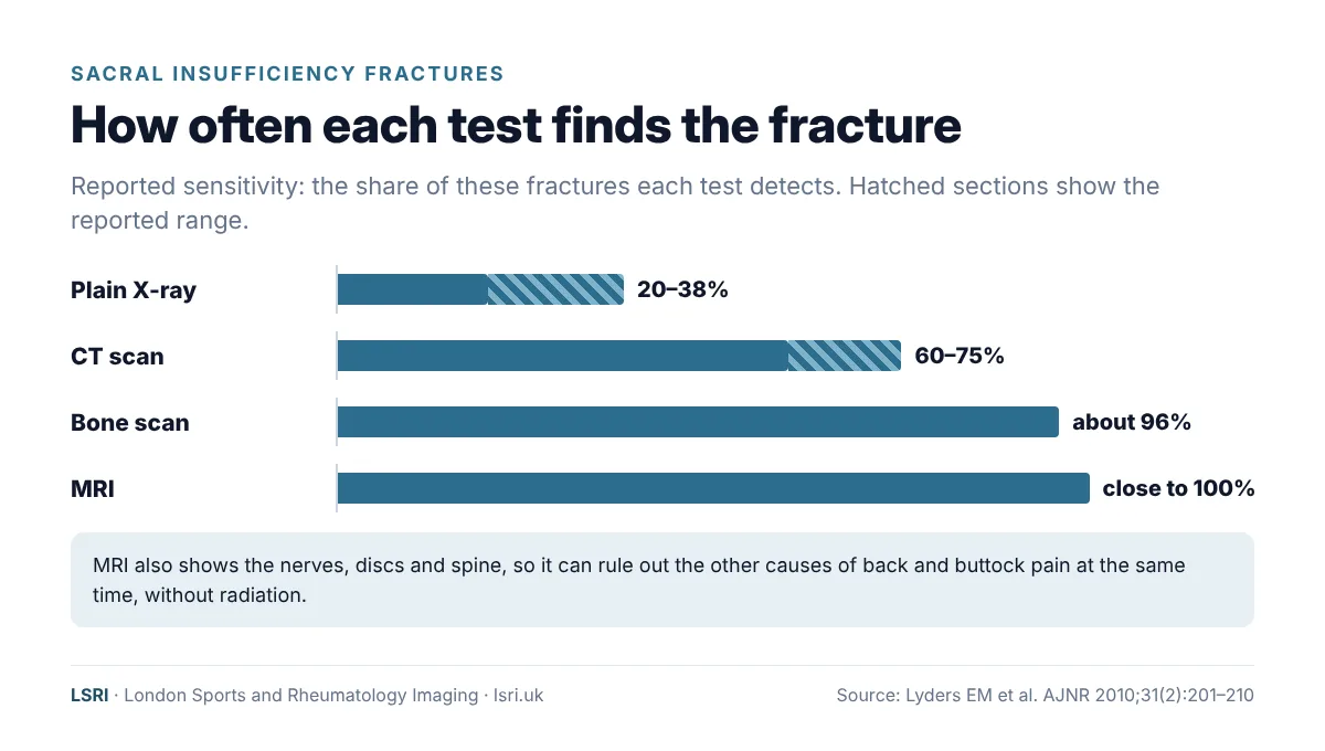
The sacrum is a curved bone, hidden behind bowel gas and the tissues of the pelvis, and the bone of someone with osteoporosis is already thin on X-ray. Together, these make sacral fractures very hard to see on a plain X-ray: reported detection rates are only about 20–38%. CT does better, at around 60–75%. MRI detects close to all of them, because it shows the swelling in the bone marrow directly.1
MRI has other advantages for someone with back and buttock pain. It uses no ionising radiation, and it also shows the spine, discs and nerves, so it can check for the other causes of the same symptoms in one visit. There is one practical point: because the pain feels like back pain, many people are first sent for an MRI of the lumbar spine. In one study, nearly two-thirds of sacral insufficiency fractures were picked up on a lumbar spine MRI, which is why the sacrum should be included in the scan and checked specifically.5 At LSRI, if the history suggests an insufficiency fracture, we tailor the scan to include the sacrum and pelvis.
Treatment of the fracture
Most sacral insufficiency fractures are treated without surgery:1
- Pain relief that allows you to move.
- Early, gentle mobilisation, with walking aids if needed. Long periods of bed rest are avoided because they weaken bones and muscles further.
- Physiotherapy to rebuild confidence, strength and balance once the diagnosis is known.
- Time: most people improve gradually over weeks to months.
For pain that does not settle, a procedure called sacroplasty, in which bone cement is injected into the fracture, is sometimes used. Fractures that make the pelvic ring unstable may need surgical fixation.1,8
A word about manipulation
Spinal and pelvic manipulation is widely used for back pain. World Health Organization guidance lists an acute fracture as a reason not to manipulate, and advises caution where bones are thinned by osteoporosis.3 This is not a criticism of manual therapy, which helps many people with back pain. It is one more reason why the diagnosis matters: once it is known, everyone treating you can adjust what they do.
The bigger picture: what the fracture says about your bones
An insufficiency fracture is a fragility fracture: a bone broken by a force that a healthy bone would withstand. It is one of the strongest signs that the skeleton is fragile, and that further fractures, including hip and spine fractures, are more likely. Treating the fracture is only half the job. The other half is protecting the rest of the skeleton.
Osteoporosis in numbers
The Royal Osteoporosis Society estimates that around 3.5 million people in the UK have osteoporosis, and that half of women and one in five men over 50 will break a bone because of it.9 An estimated 527,000 new fragility fractures occurred in the UK in 2019 alone.10 Osteoporosis is often called a silent condition, because it causes no symptoms until a bone breaks.
DEXA scans and T-scores
A DEXA (or DXA) scan measures bone mineral density, usually at the hip and lower spine. It is quick, painless, and uses a very low dose of X-rays.11 The result is given as a T-score, which compares your bone density with that of a healthy young adult.12
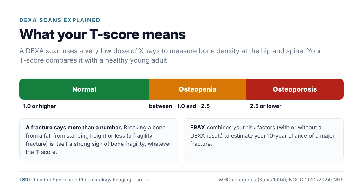
A T-score is only part of the picture. UK guidance uses a tool called FRAX, which combines risk factors such as age, sex, previous fractures, steroid use and family history, with or without a DEXA result, to estimate the chance of a major fracture over the next 10 years. That estimate guides whether treatment is recommended.4,13
What UK guidance recommends after a fragility fracture
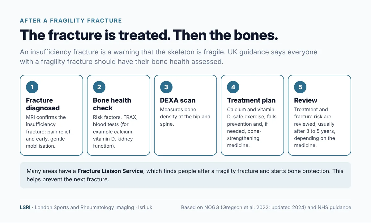
The UK clinical guideline for osteoporosis, from the National Osteoporosis Guideline Group (NOGG, updated in 2024), recommends that everyone with a fragility fracture should be assessed for osteoporosis and fracture risk, ideally through a Fracture Liaison Service.4 Many hospitals have one: a team that finds people after a fragility fracture and makes sure bone health is assessed and treated. If you have had a fragility fracture, it is reasonable to ask your GP whether a bone health assessment has been arranged.
Looking after your bones: vitamin D, calcium and exercise
Vitamin D helps the body absorb calcium. In the UK, the NHS advises that everyone should consider taking a daily supplement of 10 micrograms (400 units) of vitamin D in autumn and winter, and that some people, including those who are rarely outdoors, live in a care home or usually cover most of their skin, should take it all year round. People with dark skin, for example from an African, African-Caribbean or south Asian background, should also consider taking it all year round. The NHS also advises not taking more than 100 micrograms (4,000 units) a day unless a doctor recommends it.14
Calcium is the main mineral in bone. Adults need about 700 mg a day, which most people can get from a varied diet including dairy foods, green leafy vegetables, fortified foods and fish where you eat the bones.15 If you have osteoporosis, your doctor may recommend a calcium and vitamin D supplement alongside other treatment. Check with your GP or pharmacist before starting high-dose supplements.
Exercise is safe and recommended for people with osteoporosis. The UK consensus statement Strong, Steady and Straight recommends muscle-strengthening exercise, impact exercise suited to your fitness and fracture history, and balance training to reduce falls, along with advice on safe movement and posture.16 After a recent fracture, a physiotherapist can help you build up safely.
Falls prevention matters just as much: checking eyesight, medicines that cause dizziness, trip hazards at home and footwear can all reduce the chance of the next fracture.
Bone-strengthening medicines: alendronic acid and others
If your fracture risk is high, your doctor may recommend a medicine to strengthen your bones. The most commonly used in the UK is alendronic acid (alendronate), one of a group of medicines called bisphosphonates. It slows down the cells that break bone down, so that bone density can stabilise or improve.17
Does it work?
Yes. A 2025 Cochrane review found that, in women after the menopause who were at higher risk of fracture (for example with osteoporosis or a previous spine fracture), alendronate probably reduced spine fractures from about 5 to about 2 in every 100 women, and may also lower the risk of hip, wrist and other fractures.18
How to take it
Alendronic acid is usually taken once a week. It is absorbed poorly and can irritate the gullet, so the way you take it matters. The NHS and the patient information leaflet advise you to:17,19
- take it first thing in the morning, on an empty stomach, with a full glass of plain tap water (not mineral water, tea, coffee, juice or milk);
- swallow the tablet whole, without chewing or sucking it;
- stay upright (sitting, standing or walking) for at least 30 minutes afterwards, and do not lie down until after your first food of the day;
- wait at least 30 minutes before eating, drinking anything other than water, or taking other medicines, including calcium supplements and antacids.
Side effects
Most people take alendronic acid without problems. The most common side effects include indigestion, bloating or wind, constipation or diarrhoea, headaches, and muscle or joint pain. Stop taking it and speak to a doctor or call 111 straight away if you get heartburn or heartburn that gets worse, or pain or problems when swallowing.17
Two rare side effects are worth knowing about. Osteonecrosis of the jaw, a problem with healing of the jawbone, is rare; tell your dentist you take alendronic acid, keep up with dental check-ups, and tell your dentist and doctor straight away about a loose tooth, mouth sores, or pain or swelling in your mouth or jaw. Atypical thigh bone fractures are also rare: report any new pain in the thigh, hip or groin to your doctor.17 For most people at high fracture risk, the benefits of treatment are much greater than these rare risks.
How long for, and other options
Bisphosphonate treatment is usually reviewed after about five years of tablets (or three years of the yearly infusion), when your doctor will look at your fracture risk again and decide whether to continue or take a break.4 Other options include zoledronic acid (a yearly drip), denosumab (an injection every six months), hormone replacement therapy for some women aged 60 or under, and bone-building medicines for people at very high risk.4 Denosumab should not be stopped or delayed without a plan, because bone loss can rebound quickly and the risk of spine fractures can rise.20
Always discuss medicines with your GP or specialist. This article explains the options; it is not a recommendation for any individual.
What should you do?
- If back or buttock pain is not settling, especially if you are older, have osteoporosis, take steroids or have had pelvic radiotherapy, ask whether an insufficiency fracture has been considered.
- Ask about MRI if the cause is unclear. A normal X-ray does not rule out a sacral fracture.
- Tell anyone treating you about osteoporosis, previous fractures or steroid use, especially before manipulation.
- After the diagnosis, ask about your bones. Ask your GP for a bone health assessment: fracture risk, a DEXA scan if needed, vitamin D and calcium, exercise and falls advice, and whether a bone-strengthening medicine is right for you.
- Know the emergency signs. Back pain with numbness around the genitals or bottom, new problems controlling your bladder or bowel, or weakness in the legs needs emergency care: go to A&E or call 999.
How LSRI can help
- The right scan. When an insufficiency fracture is possible, we make sure the sacrum and pelvis are properly covered, not just the lumbar spine.
- Specialist reporting. Your MRI is reported by a consultant musculoskeletal radiologist within 24 hours. If a fracture suggests fragile bones, the report says so, so that your GP can arrange a bone health assessment.
- Clear pricing. MRI from £375 per body part, including the scan, a specialist report and access to your images.
- Time to talk. MRI with a consultation, to go through the results and next steps, is from £425.
See our private MRI scans page, read when you need an MRI for back pain, or see our case study on back pain and the sacrum.
Frequently asked questions
Can you break your sacrum without falling?
Yes. That is exactly what an insufficiency fracture is: a crack in weakened bone caused by everyday activities such as walking, standing or getting up from a chair. Many people cannot remember any injury at all.
How long does a sacral insufficiency fracture take to heal?
Most people improve gradually over several weeks to a few months, and most have little or no pain by a year. Recovery depends on general health and bone strength, which is why treating the underlying bone fragility matters.
Will an X-ray show a sacral fracture?
Often not. X-rays detect only around a fifth to just over a third of these fractures. MRI detects nearly all of them.
Do I need a DEXA scan after an insufficiency fracture?
Usually, yes, or at least a bone health assessment. UK guidance recommends that everyone with a fragility fracture is assessed for osteoporosis and fracture risk. Your GP or a Fracture Liaison Service can arrange this.
Is alendronic acid safe?
For most people at high fracture risk, yes: the benefits are much greater than the risks. Taking it correctly reduces stomach and gullet side effects. Rare side effects include jaw and thigh bone problems, so tell your dentist you take it and report new thigh, hip or groin pain. Your doctor will review treatment, usually after about five years.
Book an appointment
Call 0203 633 5040 or book online. Clinics at Harley Street (W1G 9QJ) and Harrow (HA2 7SJ). If you have emergency symptoms, go to A&E or call 999 instead.
References
- Lyders EM, Whitlow CT, Baker MD, Morris PP. Imaging and treatment of sacral insufficiency fractures. American Journal of Neuroradiology 2010;31(2):201–210. doi:10.3174/ajnr.A1666
- Sudhir G, Kalra KL, Acharya S, Chahal R. Sacral insufficiency fractures mimicking lumbar spine pathology. Asian Spine Journal 2016;10(3):558–564. doi:10.4184/asj.2016.10.3.558
- World Health Organization. WHO guidelines on basic training and safety in chiropractic. Geneva: WHO; 2005. iris.who.int
- Gregson CL, Armstrong DJ, Avgerinou C, et al. The 2024 UK clinical guideline for the prevention and treatment of osteoporosis. Archives of Osteoporosis 2025;20(1):119. doi:10.1007/s11657-025-01588-3
- Kim YY, Chung BM, Kim WT. Lumbar spine MRI versus non-lumbar imaging modalities in the diagnosis of sacral insufficiency fracture: a retrospective observational study. BMC Musculoskeletal Disorders 2018;19(1):257. doi:10.1186/s12891-018-2189-1
- Tins BJ, Garton M, Cassar-Pullicino VN, et al. Stress fracture of the pelvis and lower limbs including atypical femoral fractures: a review. Insights into Imaging 2015;6(1):97–110. doi:10.1007/s13244-014-0371-z
- Sapienza LG, Salcedo MP, Ning MS, et al. Pelvic insufficiency fractures after external beam radiation therapy for gynecologic cancers: a meta-analysis and meta-regression of 3929 patients. International Journal of Radiation Oncology, Biology, Physics 2020;106(3):475–484. doi:10.1016/j.ijrobp.2019.09.012
- Rommens PM, Hofmann A. Comprehensive classification of fragility fractures of the pelvic ring: recommendations for surgical treatment. Injury 2013;44(12):1733–1744. doi:10.1016/j.injury.2013.06.023
- Royal Osteoporosis Society. Media toolkit. theros.org.uk
- Kanis JA, Norton N, Harvey NC, et al. SCOPE 2021: a new scorecard for osteoporosis in Europe. Archives of Osteoporosis 2021;16(1):82. doi:10.1007/s11657-020-00871-9
- NHS. Bone density scan (DEXA scan). nhs.uk
- Kanis JA, Melton LJ, Christiansen C, et al. The diagnosis of osteoporosis. Journal of Bone and Mineral Research 1994;9(8):1137–1141. doi:10.1002/jbmr.5650090802
- National Osteoporosis Guideline Group (NOGG). Clinical guideline for the prevention and treatment of osteoporosis (2024 update). nogg.org.uk
- NHS. Vitamins and minerals: vitamin D. nhs.uk
- NHS. Vitamins and minerals: calcium. nhs.uk
- Brooke-Wavell K, Skelton DA, Barker KL, et al. Strong, steady and straight: UK consensus statement on physical activity and exercise for osteoporosis. British Journal of Sports Medicine 2022;56(15):837–846. doi:10.1136/bjsports-2021-104634
- NHS. Alendronic acid. nhs.uk
- Wells GA, Hsieh SC, Peterson J, et al. Alendronate for the primary and secondary prevention of osteoporotic fractures in postmenopausal women. Cochrane Database of Systematic Reviews 2025;1:CD001155. doi:10.1002/14651858.CD001155.pub3
- Alendronic Acid 70 mg tablets: patient information leaflet. Electronic medicines compendium (emc). medicines.org.uk
- Tsourdi E, Langdahl B, Cohen-Solal M, et al. Discontinuation of denosumab therapy for osteoporosis: a systematic review and position statement by ECTS. Bone 2017;105:11–17. doi:10.1016/j.bone.2017.08.003
About this case study
Patient privacy: the image is anonymised, with no names, dates or other identifying details, and non-essential details of the case have been left out. It is shared for education. Purpose: written by the LSRI clinical team to help the public recognise sacral insufficiency fractures and understand bone health, based on the research and guidance listed above. It is general information, not a diagnosis or a prescription; please discuss any medicine with your GP, specialist or pharmacist. The case is not a criticism of any practitioner: conservative care is a reasonable first step for most back pain.