Written by: LSRI Editorial Team
Medically reviewed by Dr Prashant Sankaye, Consultant MSK Radiologist ·
Last Updated: October 14, 2026
Read Time: 16 Minutes
Written by the LSRI clinical team and medically reviewed by Dr Prashant Sankaye, Consultant MSK Radiologist, on 10 October 2026.
Finding a lump under the skin is very common, and most lumps are harmless: a lipoma (a soft lump of fat), a skin cyst or a swollen gland1. Only a small number are a rare cancer called soft-tissue sarcoma, which can start in fat, muscle, blood vessels, ligaments or tendons2. These cancers are usually painless at first and can feel like an ordinary lump, so many are found late, when already large2,3.
This article explains which lumps need prompt checking, why a serious lump can be found late and why the first scan matters. We recommend having any lump scanned before it is removed or treated, usually with ultrasound first and MRI if needed. For a shorter overview, read our guide to the signs that a lump needs checking.
Key points
- Most lumps under the skin are harmless. In one hospital population, harmless (benign) soft-tissue lumps outnumbered cancerous ones by about 100 to 14.
- A lump that is getting bigger, is larger than 5 cm, lies deep, is painful or has come back after removal should be assessed promptly5,6,7.
- Smaller sarcomas at diagnosis have a better outlook. In a large UK series, the relative risk of death rose with tumour size at diagnosis8.
- Removing a sarcoma before it has been scanned and biopsied is linked to more local recurrence9,10.
- We recommend having any lump scanned before it is removed or treated, usually with ultrasound first and MRI if needed. If a scan is worrying, guidelines advise that a specialist sarcoma team plans the biopsy before any surgery3,11,12.
- Scans can suggest a sarcoma, but a biopsy examined by a pathologist confirms it3,12.
Is a lump under the skin usually harmless?
Yes. Most lumps are harmless, but a lump that is still there after two weeks, or that worries you, should be checked by a GP1.
Common harmless lumps include:
- Lipoma: a soft lump of fat that moves under the skin1.
- Skin cyst: a smooth lump under the skin1.
- Swollen glands (lymph nodes): often in the neck, armpit or groin1.
In an average general practice of 3,000 patients, at least three harmless soft-tissue tumours a year can be expected, but only about one sarcoma every 24 years4.
In the UK, sarcomas make up only about 1.4% of all cancers, around 5,300 new diagnoses a year3.
Which lumps need prompt assessment?
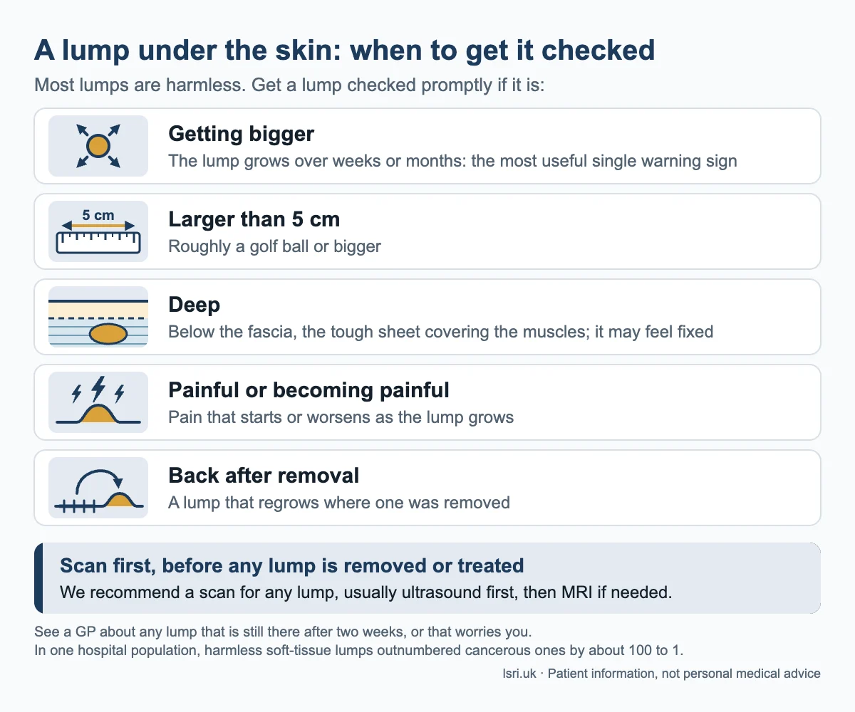
A lump should be checked promptly if it is growing, larger than about 5 cm (roughly a golf ball), deep, painful, or back after removal5,7,13.
| Warning feature | What it means | What the evidence says |
|---|---|---|
| Getting bigger | The lump grows over weeks or months | UK guidelines call increasing size the best single warning sign5 |
| Larger than 5 cm | Roughly a golf ball or bigger | Size over 5 cm was more common in cancerous lumps in a lumps clinic study of 526 people6 |
| Deep | Below the fascia, the tough sheet covering the muscles; it may feel fixed | Deeper lumps are more likely to be cancerous5,6 |
| Painful or becoming painful | Pain that starts or worsens as the lump grows | Pain was more common in cancerous than in harmless lumps6 |
| Back after removal | A lump that regrows where one was removed | Recurrent lumps are more likely to be cancerous7 |
The more of these features a lump has, the higher the risk5. But they are not perfect. UK guidelines note that, on their own, these features did not reliably separate cancerous lumps from much more common harmless ones, and only a small percentage of people referred because of them prove to have a sarcoma3. So the right response to a warning feature is a scan, not panic, and not an operation.
Why are some serious lumps found late?
Because an early sarcoma usually feels like an ordinary lump, and the route to a specialist has several steps.
- It is usually painless at first. A typical sarcoma is a solid, painless lump that is hard to move, keeps growing and may only become painful as it gets bigger2.
- It is rare. A GP practice sees far more harmless lumps than cancerous ones4.
- Deep lumps are harder to recognise, for example in the thigh3.
- An injury can seem to explain a lump. A sarcoma and a haematoma (a collection of blood, often after an injury) can look alike, even on ultrasound14. The story of the injury, such as how hard it was and whether there was bruising, needs to fit the scan14.
- The route has many steps. In a review of 76 studies, the time from first symptom to diagnosis of soft-tissue sarcoma varied from about 4 weeks to over 11 years between studies15.
Many sarcomas are large when found: UK guidelines report a typical (median) size at diagnosis of over 9 cm3.
What can delay, or the wrong first operation, cost?
Larger tumours at diagnosis do worse8, and an unplanned first removal is linked to more local recurrence and more surgery3,9,10. Evidence that a longer wait on its own worsens survival is mixed8,15.
Size at diagnosis
In a UK series of 1,460 people with a newly diagnosed bone or soft-tissue sarcoma, outcomes worsened as tumour size at diagnosis increased, even when the cancer had not spread8. For soft-tissue sarcoma that had not spread, compared with tumours of 5 cm or less, the relative risk of death over follow-up (hazard ratio) was about 2.9 times higher at 5 to 10 cm and about 8.5 times higher above 25 cm8.
The link between time and outcome is less simple. In the UK series, symptom duration did not correlate with tumour size, and people whose symptoms had lasted over a year had a slightly better outlook8. The review of 76 studies found no clinically meaningful cut-off between a short and a long delay, and conflicting results for survival in soft-tissue sarcoma, possibly because studies could not allow for how aggressive each tumour was15. This does not mean that waiting is safe. The aim is not to rush every lump, but to recognise a growing or suspicious lump and investigate it properly the first time.
The “whoops” operation
An unplanned excision, sometimes called a “whoops” procedure, is when a lump is cut out as if harmless and the laboratory then finds a sarcoma. It is reported to account for up to 40% of first operations for soft-tissue sarcoma9. Most involve lumps in or just under the skin3.
Sarcoma surgery should be planned from MRI and a biopsy taken beforehand5. After an unplanned excision, further surgery to the area where the lump was removed (re-excision) is often advised, though it is not suitable for every tumour3,10,12:
- In a combined analysis of 43 studies, tumour was still present at re-excision in 59% of patients where this was checked, and unplanned excision was linked to a 35% higher risk of local recurrence (the cancer coming back in the same place) at five years10.
- UK guidelines add that it can mean more complex surgery and more need for radiotherapy, though studies of its effect on local control conflict3.
In the combined analysis, five-year survival was not significantly worse after unplanned excision, and amputation and reconstruction rates were not higher10. But local recurrence was itself linked to worse survival10. The better path is to avoid the problem: scan first, then a biopsy by the specialist hospital team if needed, then operate once. For the wider picture, see why early imaging matters across muscle, bone and joint problems.
How should a lump be investigated?
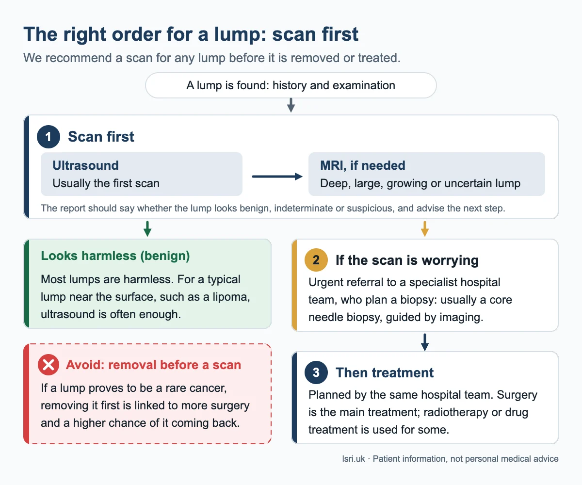
Scan first, usually with ultrasound and then MRI if needed. If the scan is worrying, a specialist hospital team plans a biopsy and then any surgery3,11,12.
Step 1: history and examination
Good practice is to record how long the lump has been there, whether it has changed in size, any symptoms and any injury, and to examine the lump, including at the time of an ultrasound scan16.
Step 2: ultrasound, usually the first scan
Ultrasound uses sound waves to make live pictures, with no radiation. UK guidelines call it an effective first test for sorting lumps3, and European guidelines recommend it first for a lump that is small (under 5 cm) and within reach of the probe11. It can confirm typical lipomas and quickly flags lumps that need more tests5,7. For a lump that is growing or larger than 5 cm, UK guidelines advise an urgent ultrasound scan or direct referral to a sarcoma diagnostic centre5.
Step 3: MRI for deep, large, growing or uncertain lumps
MRI uses a strong magnet and radio waves to show soft tissues in detail, without radiation. European guidelines call it the imaging technique of choice for lumps over 5 cm or uncertain on ultrasound, and suggest considering MRI first when cancer is suspected, a lump is deep or growing quickly, or bone or a joint is involved11. MRI should be done before a biopsy, not after, when it will help to show what the lump is11.
Step 4: a biopsy by the hospital specialist team, if needed
If imaging suggests a sarcoma, or cannot rule one out, referral to a specialist sarcoma centre should come before any biopsy11. The hospital team then arranges the standard test, a core needle biopsy: a hollow needle, usually guided by imaging, takes several thin cores of tissue3,12. They plan it so the needle track can be removed with the tumour at surgery3.
Step 5: treatment planned by the team
Surgery is the main treatment. Radiotherapy and drug treatments are used for some sarcomas, followed by regular check-ups3.
What should a scan report say?
A good report on a lump states whether it looks benign, indeterminate or suspicious for malignancy, and recommends the next step16. When features are worrying, guidelines advise urgent referral to a sarcoma service, and the report should make that recommendation clear7,11,16.
What scans cannot do
- Ultrasound depends on the operator. Its accuracy relies heavily on the skill of the person scanning3,16.
- Some lumps are indeterminate. A lump that does not look clearly harmless on ultrasound needs further imaging, usually MRI, before a firm answer7,11.
- Scans do not prove the diagnosis. Imaging suggests what a lump is; a biopsy examined by a pathologist confirms it3,12.
Does every lump need a scan?
In our view, yes: we recommend having any lump scanned and checked, usually with ultrasound first and MRI only if needed, and certainly before it is removed or treated. That does not mean every lump is a worry: most are harmless1, and for a typical lump near the surface, such as a lipoma, ultrasound is often enough5,7.
Who looks after a lump that could be serious?
A specialist sarcoma service: a hospital team that sees many of these rare cancers. Radiologists, surgeons, pathologists, oncologists and specialist nurses plan each person’s care together at a multidisciplinary team (MDT) meeting3.
UK guidelines recommend that anyone with a suspected soft-tissue sarcoma is referred to a specialist regional sarcoma service3, and European guidelines recommend early referral and treatment in specialist (reference) centres or networks12.
In a French national study of 29,497 people with a new sarcoma, surgery in a reference centre was linked to less relapse and about a 32% lower risk of death17. This was an observational study, so some of the difference may reflect which patients were treated where.
What should you do if you have a lump?
- Do not ignore a lump that is changing. See a GP if a lump lasts over two weeks, grows, is hard and does not move, is painful, red or hot, or comes back after removal1.
- Keep a simple record. Measure the lump and take a dated photo beside a ruler every few weeks. Growth is the most useful single warning sign5. You do not need to wait for several measurements before seeing a GP.
- Tell your GP about any warning feature: growing, larger than about 5 cm (roughly a golf ball), deep or fixed, or painful5,13.
- Ask for a scan before any lump is removed or treated. We recommend this for every lump, usually with ultrasound first and MRI if needed. Guidelines advise that a lump with a warning feature is assessed urgently5,7.
- Bring previous scans and reports. Earlier images show whether a lump has grown.
- If a scan is worrying, ask for referral to a specialist sarcoma centre before any biopsy. The biopsy should be planned and done by the hospital team that will treat you3,11.
- If a removed lump turns out to be a sarcoma, ask for prompt specialist referral. European guidelines advise that re-excision at a reference centre should be considered12.
How LSRI can help
LSRI is a doctor-led musculoskeletal imaging clinic. Our lumps and bumps clinic offers ultrasound-led assessment of lumps, reported by a consultant MSK radiologist. We add MRI when a lump is deep, large or uncertain on ultrasound.
- MRI from £375 per body part, including the scan, a specialist report and access to your images (from £425 with a consultation).
- Reports within 24 hours.
- Clinics at Harley Street (W1G 9QJ) and Harrow (HA2 7SJ). Phone 0203 633 5040.
- Already had a scan elsewhere? We offer a second opinion on scans and reports.
If a scan shows features that need specialist assessment, the report says so clearly and advises urgent referral to a sarcoma service. LSRI does not carry out biopsies or provide cancer treatment. If a biopsy is needed, we recommend it is done in the NHS by the hospital’s specialist sarcoma team, who will also plan any treatment.
Reading this from outside the UK? The warning features and the scan-first, biopsy-at-a-specialist-centre pathway apply everywhere and match European guidance11,12. Ask your doctor for referral to a recognised sarcoma reference centre or orthopaedic oncology service, ideally before any biopsy or surgery. European radiology guidelines also support remote second-opinion review of scans by a tumour centre when MRI findings are indeterminate or suspicious11.
Frequently asked questions
How can I tell if a lump is a harmless lipoma?
You cannot reliably tell by feel alone, which is why we recommend a scan. Lipomas are by far the most common soft-tissue lump of the limbs and trunk seen in primary care3, and ultrasound can often confirm a typical lipoma5. If imaging is unsure, a biopsy by a specialist hospital team is the way to confirm the diagnosis3,11.
Is a painless lump less worrying?
Not necessarily. Most early sarcomas are painless, and the most common presentation is a painless lump that is getting bigger2,3. Pain is a warning feature when present6. But a lack of pain is not reassuring on its own if a lump is growing, large or deep.
What size of lump should be checked?
Any lump that lasts more than two weeks, or worries you, is worth showing a GP1. A lump larger than about 5 cm, roughly a golf ball, should be assessed promptly, especially if growing or deep5,13. Smaller lumps can still be sarcomas, so growth matters as well as size5.
Is ultrasound enough, or do I need an MRI?
Ultrasound is usually the first scan, and for a small lump near the surface it can often confirm a harmless lump3,5,11. Deep, large, rapidly growing or uncertain lumps are usually assessed with MRI11.
References
- NHS. Lumps. Page last reviewed 5 February 2024. nhs.uk
- NHS. Soft tissue sarcoma: What is soft tissue sarcoma? and Symptoms. Pages last reviewed 20 February 2024. nhs.uk and nhs.uk
- Hayes AJ, Nixon IF, Strauss DC, Seddon BM, Desai A, Benson C, et al. UK guidelines for the management of soft tissue sarcomas. Br J Cancer 2025;132(1):11-31. doi:10.1038/s41416-024-02674-y
- Mannan K, Briggs TW. Soft tissue tumours of the extremities (editorial). BMJ 2005;331(7517):590. doi:10.1136/bmj.331.7517.590
- Dangoor A, Seddon B, Gerrand C, Grimer R, Whelan J, Judson I. UK guidelines for the management of soft tissue sarcomas. Clin Sarcoma Res 2016;6:20. doi:10.1186/s13569-016-0060-4
- Johnson CJ, Pynsent PB, Grimer RJ. Clinical features of soft tissue sarcomas. Ann R Coll Surg Engl 2001;83(3):203-205. PMID: 11432141
- British Sarcoma Group. Ultrasound screening of soft tissue masses in the trunk and extremity: a BSG guide for ultrasonographers and primary care. 2019. remedy.bnssg.icb.nhs.uk
- Grimer RJ. Size matters for sarcomas! Ann R Coll Surg Engl 2006;88(6):519-524. doi:10.1308/003588406X130651
- Qureshi YA, Huddy JR, Miller JD, Strauss DC, Thomas JM, Hayes AJ. Unplanned excision of soft tissue sarcoma results in increased rates of local recurrence despite full further oncological treatment. Ann Surg Oncol 2012;19(3):871-877. doi:10.1245/s10434-011-1876-z
- Larios F, Gonzalez MR, Ruiz-Arellanos K, Aquilino E Silva G, Pretell-Mazzini J. Is unplanned excision of soft tissue sarcomas associated with worse oncological outcomes? A systematic review and meta-analysis. Cancers (Basel) 2024;16(2):443. doi:10.3390/cancers16020443
- Noebauer-Huhmann IM, Vanhoenacker FM, Vilanova JC, Tagliafico AS, Weber MA, Lalam RK, et al. Soft tissue tumor imaging in adults: European Society of Musculoskeletal Radiology-Guidelines 2023: overview, and primary local imaging: how and where? Eur Radiol 2024;34(7):4427-4437. doi:10.1007/s00330-023-10425-5
- Gronchi A, Miah AB, Dei Tos AP, Abecassis N, Bajpai J, Bauer S, et al. Soft tissue and visceral sarcomas: ESMO-EURACAN-GENTURIS Clinical Practice Guidelines for diagnosis, treatment and follow-up. Ann Oncol 2021;32(11):1348-1365. doi:10.1016/j.annonc.2021.07.006
- Sarcoma UK. Do I have sarcoma? Last reviewed 2023. sarcoma.org.uk
- Berry C, Charnock M. Sarcoma or haematoma? If only it was that simple! Part 1. Ultrasound 2025;33(1):58-65. doi:10.1177/1742271X241275136
- Soomers V, Husson O, Young R, Desar I, van der Graaf W. The sarcoma diagnostic interval: a systematic review on length, contributing factors and patient outcomes. ESMO Open 2020;5(1):e000592. doi:10.1136/esmoopen-2019-000592
- The Royal College of Radiologists. Audit of ultrasound technique for the evaluation of soft tissue lumps (AuditLive template). Published 26 January 2021. rcr.ac.uk
- Blay JY, Honoré C, Stoeckle E, Meeus P, Jafari M, Gouin F, et al. Surgery in reference centers improves survival of sarcoma patients: a nationwide study. Ann Oncol 2019;30(7):1143-1153. doi:10.1093/annonc/mdz124
Book an appointment
Call 0203 633 5040 or book online. Clinics at Harley Street (W1G 9QJ) and Harrow (HA2 7SJ). If you have emergency symptoms, go to A&E or call 999 instead.
About this article
Purpose: written by the LSRI clinical team to help the public recognise which lumps under the skin need prompt checking and why a scan should come before any lump is removed or treated, based on the research and guidance listed above. It is general information, not a diagnosis. If you are worried about your own symptoms, speak to your GP or, in an emergency, go to A&E. This article does not describe any individual patient and is not a criticism of any clinician.