Written by: LSRI Editorial Team
Medically reviewed by Dr Prashant Sankaye, Consultant MSK Radiologist ·
Last Updated: October 14, 2026
Read Time: 13 Minutes
A note before you read: this article discusses anorexia nervosa and its effects on the body. If you are affected by an eating disorder, support is available. Beat, the UK’s eating disorder charity, runs helplines for England, Scotland, Wales and Northern Ireland (details at the end of this article). If you feel unsafe right now, call 999 or go to A&E.
Anorexia nervosa is a serious mental illness that affects the whole body, including the skeleton. One of its lesser-known effects is on the bone marrow, the soft tissue inside our bones that makes blood cells and stores fat. When the body is severely undernourished, the marrow can undergo gelatinous transformation: its fat cells and blood-forming cells shrink and are replaced by a jelly-like substance. This change cannot be seen or felt from the outside, but it has a distinctive appearance on MRI. Recognising it matters, for patients, for families and for the clinicians caring for them. Using an anonymised case from our clinic, this article explains what gelatinous marrow transformation is, how MRI shows it, why it matters for bone health, and where to find support.
Key points
- Gelatinous marrow transformation, also called serous atrophy of bone marrow, is a change in the bone marrow caused by severe undernutrition. Anorexia nervosa is one of its most common causes.1,2
- In one study of people with anorexia nervosa who had a bone marrow examination, about half showed gelatinous change.3
- On MRI, marrow that should look bright on T1 images because of its fat turns dark, and looks bright on fluid-sensitive images. It can be mistaken for swelling, infection, a tumour or even a technical fault.2,4
- It often goes hand in hand with low bone density and a higher risk of fractures, including stress fractures that can be hard to see.4,5,6,7
- These changes usually improve with nutritional recovery and specialist treatment. Recovery from anorexia nervosa is possible, and help is available.
The case: what MRI showed in the thigh bone
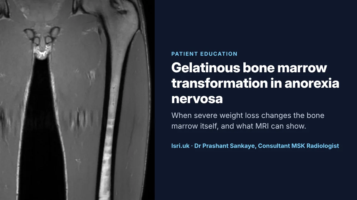
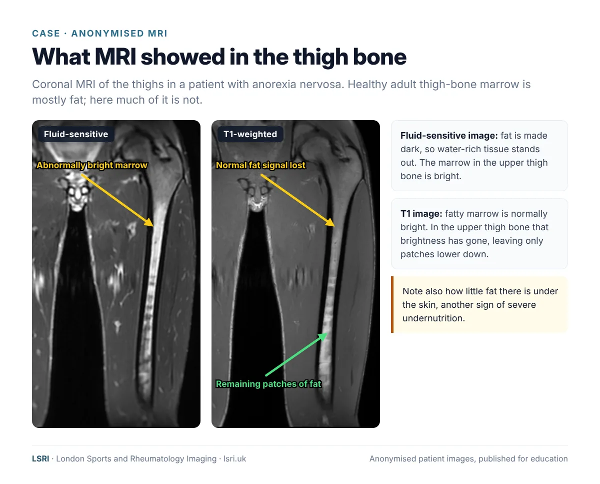
An adult patient with anorexia nervosa had an MRI scan of the thighs at LSRI. The scan showed that much of the normal fatty marrow in the upper part of the thigh bone (femur) had changed. On T1-weighted images the marrow had lost its normal brightness, with only patches of fat remaining lower down the bone. On fluid-sensitive images the same marrow was abnormally bright. There was also very little fat under the skin. Together, these are the typical appearances of gelatinous transformation of the bone marrow.
What is bone marrow, and what changes?
Bone marrow comes in two main kinds. Red marrow makes red blood cells, white blood cells and platelets. Yellow marrow is mostly fat. In childhood, most marrow is red. Through adolescence and adulthood, much of it converts to fatty yellow marrow, especially in the long bones of the arms and legs. In a healthy adult, the shaft of the thigh bone is mostly fat.
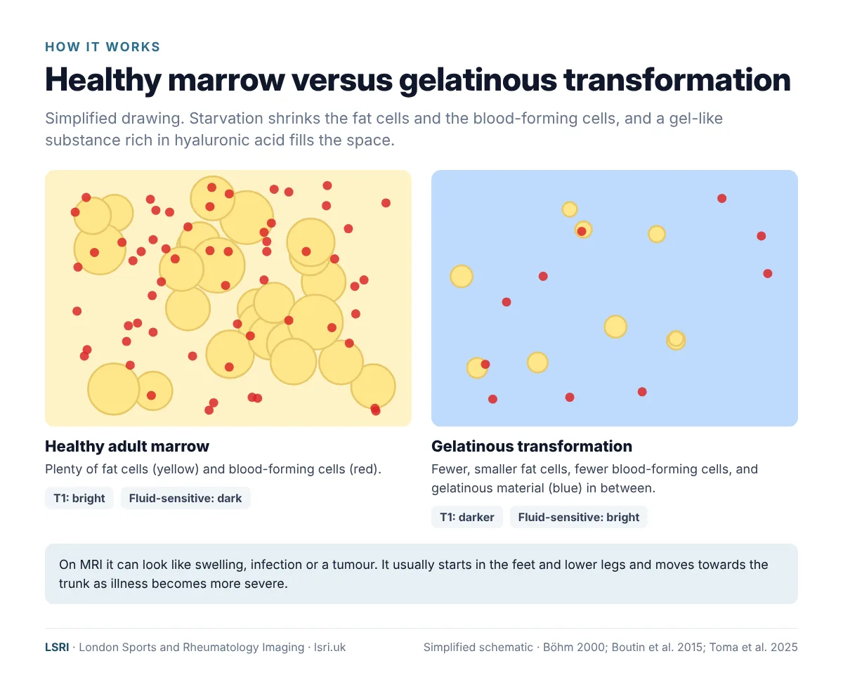
When the body does not get enough energy from food over a long period, it starts to use its reserves, including the fat stored in the bone marrow. Under the microscope, gelatinous transformation has three features:1,8
- the fat cells in the marrow shrink;
- the blood-forming cells become fewer (the marrow becomes “hypoplastic”);
- a gel-like material, rich in a substance called hyaluronic acid, builds up in the spaces between the cells.
Gelatinous transformation is not unique to anorexia nervosa. It can happen with any cause of severe, prolonged undernutrition or wasting, including advanced cancer, serious chronic illness, some infections and alcohol-related illness.1,8 In younger people, anorexia nervosa is one of the most common causes.1,4
The change is usually most marked furthest from the trunk, in the bones of the feet and lower legs. In more severe illness it is also seen in the hips and spine. Early MRI studies found that the extent of marrow change was linked to the severity of the illness.9,10
What it looks like on MRI
MRI is particularly good at showing bone marrow because it can tell fat and water apart:
- On T1-weighted images, fat is bright. Healthy adult marrow in the limbs is therefore bright. With gelatinous transformation, the marrow turns darker, because the fat has gone.
- On fluid-sensitive images, such as STIR or fat-suppressed T2, fat is made dark and water stands out. The water-rich gelatinous material looks bright.
In one of the larger imaging studies, the fat under the skin was also abnormal in almost every patient with these marrow changes, a useful clue for the radiologist.4 The same study found fractures in almost half of the patients.
Why it can be misread
Bright marrow on a fluid-sensitive scan has many possible causes, including injury, infection, inflammation and tumours. Gelatinous transformation can also be mistaken for a technical problem with the scan: in one study, some scans had been repeated because the appearance was put down to a technical fault with the scan, such as failed fat suppression.2,4 Recognising the pattern matters because it can prevent unnecessary repeat scans and other investigations, and point care in the right direction.2
The marrow change can also hide a stress fracture, because the swelling that normally makes a stress fracture stand out on MRI blends in with the abnormal marrow around it. Radiologists who know this look especially carefully for fracture lines.7 Occasionally, the marrow appearance on a scan is the first clue that someone may be seriously undernourished, which is why a careful, sensitive report matters.7
Why MRI matters in anorexia nervosa
- Bone pain should never be dismissed. People with anorexia nervosa have a higher risk of fractures.6,11 In one MRI study of people with these marrow changes, almost half had fractures, mostly in the hips and legs.4 X-rays are often normal in the early weeks of a stress fracture.12
- MRI shows the whole picture. It shows marrow change, bone stress injuries and soft-tissue problems in one scan, without ionising radiation. That matters for young people and for anyone who may need repeat imaging.
- The right interpretation avoids harm. A specialist musculoskeletal radiologist who recognises gelatinous transformation can avoid a mistaken diagnosis of infection or tumour, and can flag what the scan means for the person’s wider care.
- It is not the same as a bone density scan. A DEXA scan measures bone density and fracture risk. MRI shows the structure of the bone and marrow. Your care team may recommend one, the other, or both.
Effects on bone and blood
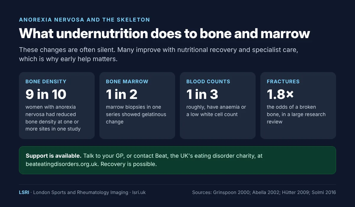
Bone density. Anorexia nervosa is a well-recognised cause of low bone density in young women. In one study of women with anorexia nervosa, more than 9 in 10 had reduced bone density in at least one part of the skeleton, and almost 4 in 10 were in the osteoporosis range at one or more sites.5 This matters because peak bone mass is built in the teens and twenties: bone that is not built then can be hard to make up later.
Fractures. A large review that pooled research on eating disorders found that people with anorexia nervosa had nearly twice the odds of a fracture.6 Girls and young women with anorexia nervosa also have a higher fracture risk than their peers, even when their bone density scan looks normal.11
Blood counts. Because the marrow makes blood cells, roughly a third of people with anorexia nervosa have anaemia or a low white blood cell count, and some have low platelets.13 In people with severe illness, these changes are common and usually improve with nutritional rehabilitation.14
Looking well is not the same as being well
The Royal College of Psychiatrists’ guidance on medical emergencies in eating disorders makes an important point for families and clinicians alike: “Patients can appear well and this can falsely reassure the clinician.”15 Normal blood tests or a normal appearance do not mean the body, including the skeleton, has not been affected.
Can it get better?
Yes. Gelatinous marrow transformation is a response to undernutrition, and it usually improves with nutritional recovery and specialist treatment, although this can take time.1,2 Blood counts usually recover too.14 Bone density tends to be slower to recover, which is one more reason why early help makes a difference. Recovery from anorexia nervosa is possible.
For families, friends, coaches and teachers
If you are worried about someone, you do not need to be certain before you act. The NHS describes common signs of anorexia nervosa, including missing meals or eating very little, exercising excessively and an overwhelming fear of gaining weight, along with physical signs such as feeling cold, dizzy or very tired, and periods stopping.16 Bone pain, or a stress fracture in someone who has lost weight, is another reason to raise the question gently.
- Talk to the person privately and kindly, focusing on how they are feeling rather than on food or weight.
- Encourage them to see their GP. Early treatment gives the best chance of recovery.
- Look after yourself too. Beat has support for families and friends as well as for people with eating disorders.
Where to get help
Support for eating disorders
- Your GP is usually the first step to getting assessed and referred to a specialist eating disorder service.
- Beat helplines: England 0808 801 0677 · Scotland 0808 801 0432 · Wales 0808 801 0433 · Northern Ireland 0808 801 0434. Opening hours and web chat are on the Beat website.17
- If you need urgent help, call NHS 111. Beat is not a crisis service: in an emergency, call 999 or go to A&E.
- If you need to talk to someone now, Samaritans are available 24 hours a day on 116 123, or you can text SHOUT to 85258.
How LSRI can help
LSRI is a specialist imaging clinic, not an eating disorder service. If you have, or think you may have, an eating disorder, your GP and a specialist eating disorder team are the right people to lead your care. Where imaging is needed, for example for bone pain or a suspected stress fracture, we can help:
- Specialist reporting. Your MRI is reported by a consultant musculoskeletal radiologist within 24 hours, with careful attention to the bone marrow as well as to fractures.
- Working with your team. We share results with your GP or specialist team, with your agreement, so that the scan informs your wider care.
- Second opinions. Clinicians and patients can ask for a specialist second opinion on an existing MRI.
- Time to talk. MRI with a consultation, to go through the results and next steps, is from £425.
See our private MRI scans page, or read our guides to the female athlete triad and REDs and hip stress fractures.
Frequently asked questions
Is gelatinous marrow transformation dangerous?
The marrow change itself is a sign of how severely the body has been affected by undernutrition, rather than a separate disease. It matters because it goes with reduced blood cell production, weaker bones and a higher risk of fractures. It is a reason to make sure specialist care is in place.
Is it reversible?
Usually, yes. The marrow generally recovers with nutritional rehabilitation, although this can take time. Bone density recovers more slowly, which is why early treatment matters.
Can a blood test show it?
Not directly. Blood tests may show low blood counts, which can go with marrow change, but they can also be normal. MRI or a bone marrow examination shows the marrow itself.
Does it only happen in anorexia nervosa?
No. It can happen with any cause of severe, prolonged undernutrition or wasting, including advanced cancer, serious chronic illness and some infections.
My scan report says “serous atrophy”. Is that the same thing?
Yes. Serous atrophy of bone marrow, gelatinous transformation and gelatinous marrow are different names for the same change. If anything in your report worries you, ask the doctor who requested the scan to go through it with you.
Book an appointment
Call 0203 633 5040 or book online. Clinics at Harley Street (W1G 9QJ) and Harrow (HA2 7SJ).
References
- Böhm J. Gelatinous transformation of the bone marrow: the spectrum of underlying diseases. American Journal of Surgical Pathology 2000;24(1):56–65. doi:10.1097/00000478-200001000-00007
- Toma T, Viegas S, Amiras D, Lord CJ. Recognising serous atrophy of bone marrow: a review of imaging findings. Skeletal Radiology 2025;54(12):2597–2606. doi:10.1007/s00256-025-04963-w
- Abella E, Feliu E, Granada I, et al. Bone marrow changes in anorexia nervosa are correlated with the amount of weight loss and not with other clinical findings. American Journal of Clinical Pathology 2002;118(4):582–588. doi:10.1309/2Y7X-YDXK-006B-XLT2
- Boutin RD, White LM, Laor T, et al. MRI findings of serous atrophy of bone marrow and associated complications. European Radiology 2015;25(9):2771–2778. doi:10.1007/s00330-015-3692-5
- Grinspoon S, Thomas E, Pitts S, et al. Prevalence and predictive factors for regional osteopenia in women with anorexia nervosa. Annals of Internal Medicine 2000;133(10):790–794. doi:10.7326/0003-4819-133-10-200011210-00011
- Solmi M, Veronese N, Correll CU, et al. Bone mineral density, osteoporosis, and fractures among people with eating disorders: a systematic review and meta-analysis. Acta Psychiatrica Scandinavica 2016;133(5):341–351. doi:10.1111/acps.12556
- Tins B, Cassar-Pullicino V. Marrow changes in anorexia nervosa masking the presence of stress fractures on MR imaging. Skeletal Radiology 2006;35(11):857–860. doi:10.1007/s00256-005-0053-5
- Shergill KK, Shergill GS, Pillai HJ. Gelatinous transformation of bone marrow: rare or underdiagnosed? Autopsy and Case Reports 2017;7(4):8–17. doi:10.4322/acr.2017.039
- Vande Berg BC, Malghem J, Devuyst O, et al. Anorexia nervosa: correlation between MR appearance of bone marrow and severity of disease. Radiology 1994;193(3):859–864. doi:10.1148/radiology.193.3.7972838
- Vande Berg BC, Malghem J, Lecouvet FE, et al. Distribution of serouslike bone marrow changes in the lower limbs of patients with anorexia nervosa: predominant involvement of the distal extremities. American Journal of Roentgenology 1996;166(3):621–625. doi:10.2214/ajr.166.3.8623638
- Faje AT, Fazeli PK, Miller KK, et al. Fracture risk and areal bone mineral density in adolescent females with anorexia nervosa. International Journal of Eating Disorders 2014;47(5):458–466. doi:10.1002/eat.22248
- Wright AA, Hegedus EJ, Lenchik L, et al. Diagnostic accuracy of various imaging modalities for suspected lower extremity stress fractures: a systematic review with evidence-based recommendations for clinical practice. American Journal of Sports Medicine 2016;44(1):255–263. doi:10.1177/0363546515574066
- Hütter G, Ganepola S, Hofmann WK. The hematology of anorexia nervosa. International Journal of Eating Disorders 2009;42(4):293–300. doi:10.1002/eat.20610
- Sabel AL, Gaudiani JL, Statland B, Mehler PS. Hematological abnormalities in severe anorexia nervosa. Annals of Hematology 2013;92(5):605–613. doi:10.1007/s00277-013-1672-x
- Royal College of Psychiatrists. Medical emergencies in eating disorders (MEED): guidance on recognition and management (CR233). May 2022, updated December 2025. rcpsych.ac.uk
- NHS. Anorexia nervosa: symptoms. nhs.uk
- Beat. Helplines and support. beateatingdisorders.org.uk
About this article
Patient privacy: the images are anonymised, with no names, dates or other identifying details, and non-essential details of the case have been left out. They are shared for education. Purpose: written by the LSRI clinical team to explain a bone marrow change that is not widely known, based on the research and guidance listed above. It is general information, not a diagnosis or treatment advice. Eating disorders are treated by GPs and specialist eating disorder services; if you are worried about yourself or someone else, start with your GP or Beat.