Written by: LSRI Editorial Team
Medically reviewed by Dr Prashant Sankaye, Consultant MSK Radiologist ·
Last Updated: October 14, 2026
Read Time: 14 Minutes
Most hip and groin pain comes from a strained muscle or an irritated tendon, and it settles with time and a good physiotherapy programme. But a small number of people whose hip pain “just won’t settle” have something different: a stress fracture of the femoral neck, the short bridge of bone just below the ball of the hip joint. These fractures are uncommon, easy to miss on an X-ray and important to catch early, because if the bone goes on to break completely the consequences can be serious and long-lasting. This anonymised case from our clinic, in which more than a month of hip pain treated with physiotherapy turned out to be a fracture, shows the warning signs and explains why MRI is the test that answers the question.
Key points
- Femoral neck stress fractures are uncommon, at around 3% of stress fractures in athletes, but they are among the most important not to miss.1
- The typical symptom is a deep ache in the groin or the front of the hip that comes on with activity, often with pain at the end of hip movement and sometimes a limp.
- X-rays are often normal in the first weeks. MRI is the most accurate test and can show a bone stress injury before a crack is visible.2,3
- Fractures on the top (tension) side of the femoral neck, and any that have displaced, usually need urgent surgery.1,4
- If hip pain is not improving as expected, or is getting worse, ask whether a stress fracture has been ruled out. If you cannot put weight on the leg, go to A&E.
The case: more than a month of hip pain
An adult patient developed pain in the left hip. For more than a month it was treated as a soft-tissue problem with physiotherapy. That was a sensible place to start: most hip pain is muscular or tendon-related, and it usually improves with a well-planned rehabilitation programme.
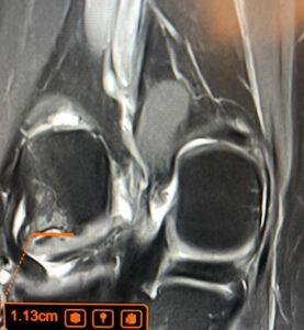
But this pain did not settle. An MRI scan at LSRI showed a stress fracture of the femoral neck on the left, on the compression side (the lower, inner edge of the bone), with extensive swelling (oedema) in the surrounding bone marrow and fluid in the hip joint. Compression-side fractures are the more common type in young, active people and, if they are small and caught early, many heal without surgery.1,4
This case is not about blame. In its early stages a femoral neck stress fracture can feel very like a groin strain or a hip tendon problem, and a first X-ray may well be normal. The lesson is about timing: when pain does not follow the expected course, the diagnosis needs to be looked at again, and that sometimes means a scan.
What the MRI showed
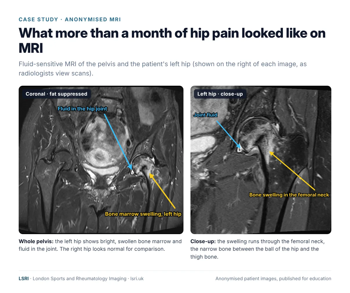
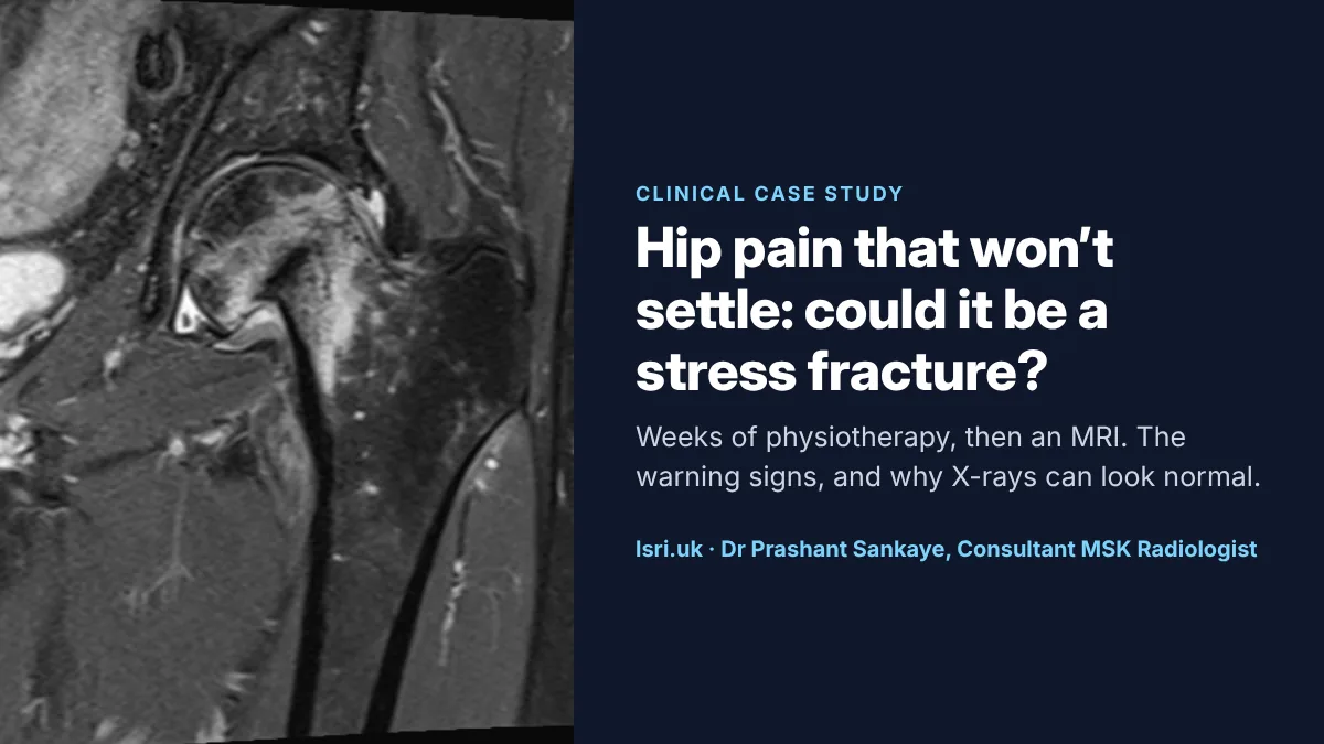
These images use a fat-suppressed (fluid-sensitive) sequence. It darkens fat, so anything containing extra water, such as swelling inside a bone, shows up bright. In the left hip, the bone marrow of the femoral neck is bright and swollen, and there is a small amount of fluid in the joint. The right hip looks normal and gives a useful comparison.
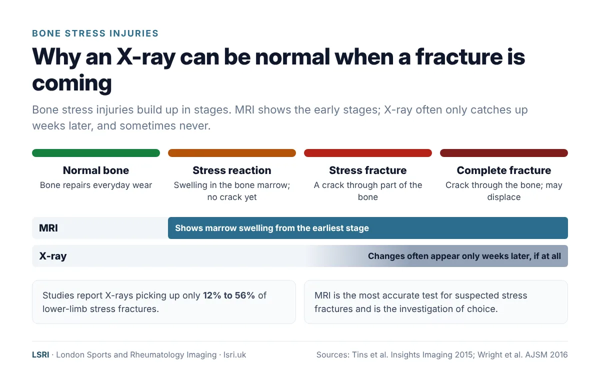
On MRI, the earliest sign of a bone stress injury is swelling in the bone marrow, before any crack appears. As the injury progresses, a fracture line develops, usually seen as a thin dark line running into the bone from its outer shell (the cortex). MRI can grade how far along this process an injury is, and higher grades generally take longer to recover from.5,6 That grading helps doctors plan treatment and give a realistic timescale for returning to sport or work.
What is a femoral neck stress fracture?
Bone is living tissue. It constantly repairs the tiny amounts of damage caused by everyday activity, and it gets stronger in response to load. A stress fracture develops when bone is loaded repeatedly, faster than it can repair itself, until the accumulated damage becomes a crack.
There are two broad types:3
- Fatigue fractures: abnormal or excessive load on normal bone. These are typical in runners, endurance athletes and military recruits, especially after a sudden increase in training.
- Insufficiency fractures: normal, everyday load on bone that has been weakened, for example by osteoporosis. Our article on sacral insufficiency fractures explains these in more detail.
The femoral neck carries the whole weight of the body with every step, and much more when running or jumping. Its upper (tension) side is one of a handful of “high-risk” stress fracture sites: places where a fracture has a higher chance of progressing, healing poorly or needing surgery.7,8 Fractures on the lower (compression) side are lower risk, but any femoral neck stress fracture can progress, so all need careful specialist management.1,4
Why the side of the bone matters
Where the fracture sits on the femoral neck changes how urgently it needs treating.1,9
- Compression-side fractures start on the lower, inner edge of the femoral neck. They are the more common type in young athletes. If they are small and caught early, many heal with a period of protected weight bearing (often on crutches) and careful monitoring. Larger ones may need surgery.
- Tension-side fractures start on the upper, outer edge. Here the forces tend to pull the crack open, so the risk of the fracture spreading across the bone is higher. These usually need urgent surgical fixation with screws.4
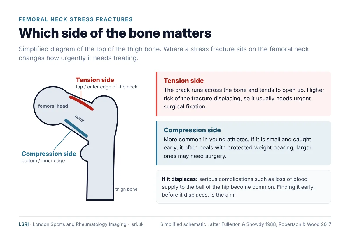
If a femoral neck stress fracture displaces (the two pieces of bone shift apart), it becomes an orthopaedic emergency. Complications are then common, including loss of blood supply to the ball of the hip (avascular necrosis) and failure of the fracture to heal. Reviews of the research report avascular necrosis in roughly a quarter to two-fifths of displaced fractures.1 You can read more about avascular necrosis of the hip in our guide to how avascular necrosis of the hip is diagnosed.
Why delay matters
The main reason to look hard for these fractures is that serious complications are much more likely once a fracture has displaced, so finding it early, while it is still incomplete, matters.1,4 In a classic study of 23 athletes with femoral neck stress fractures, the diagnosis took an average of 14 weeks from the first symptoms. Almost a third (30%) went on to have complications needing major surgery, most of them in fractures that had displaced, and all the elite athletes in the study had to end their sporting careers.10
Symptoms and warning signs
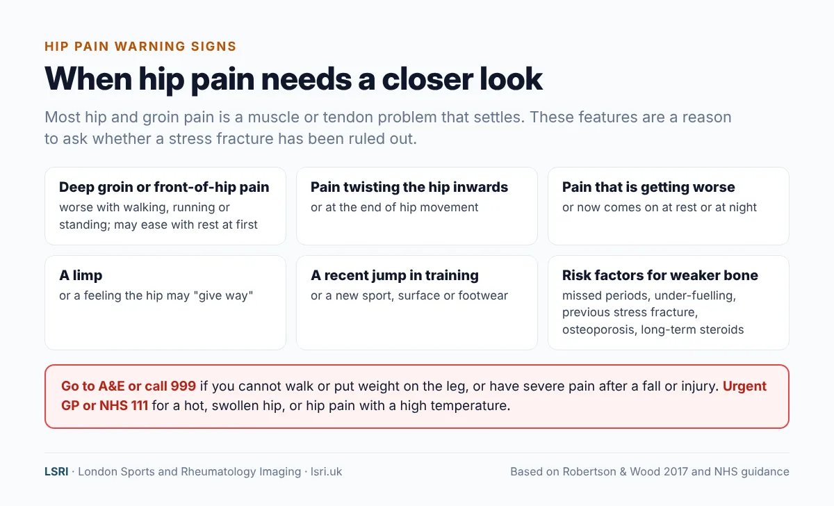
The most common symptom is pain at the front of the hip or deep in the groin, brought on by weight-bearing activity such as walking or running. At first it may ease with rest; later it may come on sooner, last longer, or be present at night.1 Pain at the end of hip movement, especially when the hip is turned inwards, is a common finding on examination.
Some clinicians ask patients to hop on the painful leg, because pain on hopping can be a clue. But this test has not been properly validated, and a negative result does not rule out a fracture.11 It is not a test to try on yourself if you are worried: if a femoral neck stress fracture is present, extra load is the last thing it needs.
Who is at higher risk?
- A recent jump in training: more distance, more speed work, a new sport, a new surface or new footwear.
- Low energy availability: not eating enough to match the energy used in training, with or without an eating disorder. In women, missed or irregular periods are an important warning sign. Our article on the female athlete triad and REDs explains why. Men can be affected too.12
- Weaker bones: low bone density or osteoporosis, long-term steroid treatment, or a previous stress fracture.1,3
Why MRI is the right test
X-rays are often normal early on. A systematic review found that X-rays detected only between 12% and 56% of lower-limb stress fractures.2 Changes on X-ray typically lag behind symptoms by two to three weeks or more.3 For the femoral neck specifically, up to two-thirds of stress fractures have a normal X-ray when the patient first presents, and around half never show on X-ray at all.1 A normal X-ray is therefore not reassurance on its own.
MRI is the most accurate test. The same review found MRI to be the most sensitive and specific test for suspected stress fractures, and expert reviews describe it as the investigation of choice.2,3 Early research in endurance athletes with hip pain found MRI better than bone scanning for working out the cause.13 MRI has other advantages too:
- It shows the early “stress reaction” stage, so treatment can start before a crack forms.
- It grades the injury and shows whether the fracture is on the tension or compression side, which guides treatment.
- It uses no ionising radiation, which matters for young people and anyone who may need repeat scans.
- It shows the other causes of hip and groin pain, such as tendon and muscle injuries, avascular necrosis and early arthritis, in the same scan.
What happens after diagnosis?
Because a femoral neck stress fracture can progress to a complete break, it should be assessed promptly by an orthopaedic surgeon, and urgently if it is on the tension side or has displaced.1,4,7,8 Treatment depends on the type and size of the fracture:1,4
- Small compression-side fractures: stopping running and impact activity, crutches to protect the hip for a period, and close follow-up, often with repeat imaging. Return to sport is gradual and guided by symptoms and the specialist team.
- Tension-side, larger or displaced fractures: usually surgical fixation.
Just as important is finding out why the fracture happened: the training load, footwear and surfaces; energy availability and nutrition; menstrual health; vitamin D; and, where appropriate, a bone density (DEXA) scan. Without that, the same thing can happen again.
What should you do if your hip pain isn’t settling?
- Notice the pattern. Pain that is getting worse rather than better, that comes on sooner with activity, or that is now present at rest or at night, is not following the expected course.
- Reduce the load. Don’t push through groin or hip pain that is getting worse with running or long walks.
- Ask for your diagnosis to be reviewed. It is reasonable to ask your physiotherapist or GP directly: “Has a stress fracture been ruled out?”
- Ask about MRI if the cause is still unclear. If an X-ray is normal but the symptoms fit, MRI is the test that answers the question.
- Know when it is an emergency. If you cannot walk or put weight on the leg, or you have severe pain after a fall or injury, go to A&E or call 999. A hot, swollen hip, or hip pain with a high temperature, needs urgent advice from your GP or NHS 111.14
How LSRI can help
- Advice before you book. Call us to discuss whether a scan is likely to help, and which one. If you have emergency symptoms, we will direct you to A&E.
- Specialist reporting. Your MRI is reported by a consultant musculoskeletal radiologist within 24 hours. If the scan shows a fracture that needs urgent treatment, we contact you and your GP so that the next steps are not delayed.
- Clear pricing. MRI from £375 per body part, including the scan, a specialist report and access to your images.
- Time to talk. MRI with a consultation, to go through the results and next steps, is from £425.
See our private MRI scans page, our guide on stress fractures that don’t show on X-ray, read when runners need an MRI, or see our case study on a heel bone fracture found after eight months of pain.
Frequently asked questions
Will a hip stress fracture show on an X-ray?
Often not at first. X-ray changes usually lag behind symptoms by weeks, and many stress fractures are never visible on X-ray. A normal X-ray does not rule out a stress fracture if the symptoms fit. MRI is the most accurate test.
Is it a groin strain or a stress fracture?
They can feel similar. A strain usually follows a specific moment of injury and steadily improves. Stress fracture pain tends to build up with weight-bearing activity, gets worse rather than better if training continues, and may come on at rest or at night. If you are not sure, ask for a review rather than training through it.
How long does a femoral neck stress fracture take to heal?
It depends on the type and severity. Many stress fractures heal in roughly 4 to 12 weeks with the right management,11 but femoral neck fractures are managed more cautiously, and a full return to running can take several months. Fractures that need surgery take longer. Your specialist team will guide this, often using repeat imaging.
Can I keep walking on a hip stress fracture?
Keep it to a minimum until it has been assessed. If a femoral neck stress fracture is suspected, you will usually be advised to stop running and impact activity and may be given crutches until a specialist has seen you. If you cannot put weight on the leg at all, go to A&E.
Do I need a GP referral for a private hip MRI?
No GP referral is needed in most cases if you are paying for yourself. If you are claiming through private medical insurance, your insurer may require a referral, so check with them first. We always recommend sharing your results with your GP.
Book an appointment
Call 0203 633 5040 or book online. Clinics at Harley Street (W1G 9QJ) and Harrow (HA2 7SJ). If you cannot put weight on your leg, or have emergency symptoms, go to A&E or call 999 instead.
References
- Robertson GA, Wood AM. Femoral neck stress fractures in sport: a current concepts review. Sports Medicine International Open 2017;1(2):E58–E68. doi:10.1055/s-0043-103946
- Wright AA, Hegedus EJ, Lenchik L, et al. Diagnostic accuracy of various imaging modalities for suspected lower extremity stress fractures: a systematic review with evidence-based recommendations for clinical practice. American Journal of Sports Medicine 2016;44(1):255–263. doi:10.1177/0363546515574066
- Tins BJ, Garton M, Cassar-Pullicino VN, et al. Stress fracture of the pelvis and lower limbs including atypical femoral fractures: a review. Insights into Imaging 2015;6(1):97–110. doi:10.1007/s13244-014-0371-z
- Bernstein EM, Kelsey TJ, Cochran GK, et al. Femoral neck stress fractures: an updated review. Journal of the American Academy of Orthopaedic Surgeons 2022;30(7):302–311. doi:10.5435/JAAOS-D-21-00398
- Nattiv A, Kennedy G, Barrack MT, et al. Correlation of MRI grading of bone stress injuries with clinical risk factors and return to play: a 5-year prospective study in collegiate track and field athletes. American Journal of Sports Medicine 2013;41(8):1930–1941. doi:10.1177/0363546513490645
- Hoenig T, Tenforde AS, Strahl A, et al. Does magnetic resonance imaging grading correlate with return to sports after bone stress injuries? A systematic review and meta-analysis. American Journal of Sports Medicine 2022;50(3):834–844. doi:10.1177/0363546521993807
- Kaeding CC, Yu JR, Wright R, et al. Management and return to play of stress fractures. Clinical Journal of Sport Medicine 2005;15(6):442–447. doi:10.1097/01.jsm.0000188207.62608.35
- McInnis KC, Ramey LN. High-risk stress fractures: diagnosis and management. PM&R 2016;8(3 Suppl):S113–S124. doi:10.1016/j.pmrj.2015.09.019
- Fullerton LR Jr, Snowdy HA. Femoral neck stress fractures. American Journal of Sports Medicine 1988;16(4):365–377. doi:10.1177/036354658801600411
- Johansson C, Ekenman I, Törnkvist H, Eriksson E. Stress fractures of the femoral neck in athletes: the consequence of a delay in diagnosis. American Journal of Sports Medicine 1990;18(5):524–528. doi:10.1177/036354659001800514
- Patel DS, Roth M, Kapil N. Stress fractures: diagnosis, treatment, and prevention. American Family Physician 2011;83(1):39–46. aafp.org
- Mountjoy M, Ackerman KE, Bailey DM, et al. 2023 International Olympic Committee’s (IOC) consensus statement on Relative Energy Deficiency in Sport (REDs). British Journal of Sports Medicine 2023;57(17):1073–1098. doi:10.1136/bjsports-2023-106994
- Shin AY, Morin WD, et al. The superiority of magnetic resonance imaging in differentiating the cause of hip pain in endurance athletes. American Journal of Sports Medicine 1996;24(2):168–176. doi:10.1177/036354659602400209
- NHS. Hip pain in adults. nhs.uk
About this case study
Patient privacy: the images are anonymised, with no names, dates or other identifying details, and non-essential details of the case have been left out. They are shared for education. Purpose: written by the LSRI clinical team to help the public recognise when hip pain needs a closer look, based on the research and guidance listed above. It is general information, not a diagnosis; if you are worried about your own symptoms, speak to your GP or, in an emergency, go to A&E. The case is not a criticism of any clinician: physiotherapy is the usual and appropriate first step for most hip pain.