Written by: LSRI Editorial Team
Medically reviewed by Dr Prashant Sankaye, Consultant MSK Radiologist ·
Last Updated: October 14, 2026
Read Time: 12 Minutes
Heel pain is common, and most of it is plantar fasciitis: irritation of the thick band of tissue under the foot, which usually improves with time and simple measures such as stretching, taping and supportive footwear. So when someone has heel pain, it is natural to treat it as plantar fasciitis. But not all heel pain is plantar fasciitis. This anonymised case from our clinic, in which eight months of heel pain and physiotherapy ended with an MRI showing a fracture of the heel bone, shows why heel pain that is not improving as expected should have its diagnosis reviewed. It also explains the principle at the centre of how we work at LSRI: in musculoskeletal medicine, diagnose first, then treat.
Key points
- Plantar fasciitis is the most common cause of heel pain, but fractures of the heel bone (calcaneus) can cause similar symptoms.1
- Calcaneal fractures can be missed in two main ways: stress fractures that do not show on early X-rays, and subtle fractures, for example of the front part of the heel bone, that are easily overlooked or mistaken for a sprain.2,3,4
- Pain when the sides of the heel are squeezed (the calcaneal squeeze test) suggests a calcaneal stress fracture.1
- MRI shows both the fracture and the swelling in the bone around it, even when X-rays are normal.5,6
- Most musculoskeletal pain does not need a scan. But international guidance accepts imaging when a serious cause is suspected, when pain is not responding to treatment as expected, or when the result is likely to change treatment.7
The case: eight months of heel pain
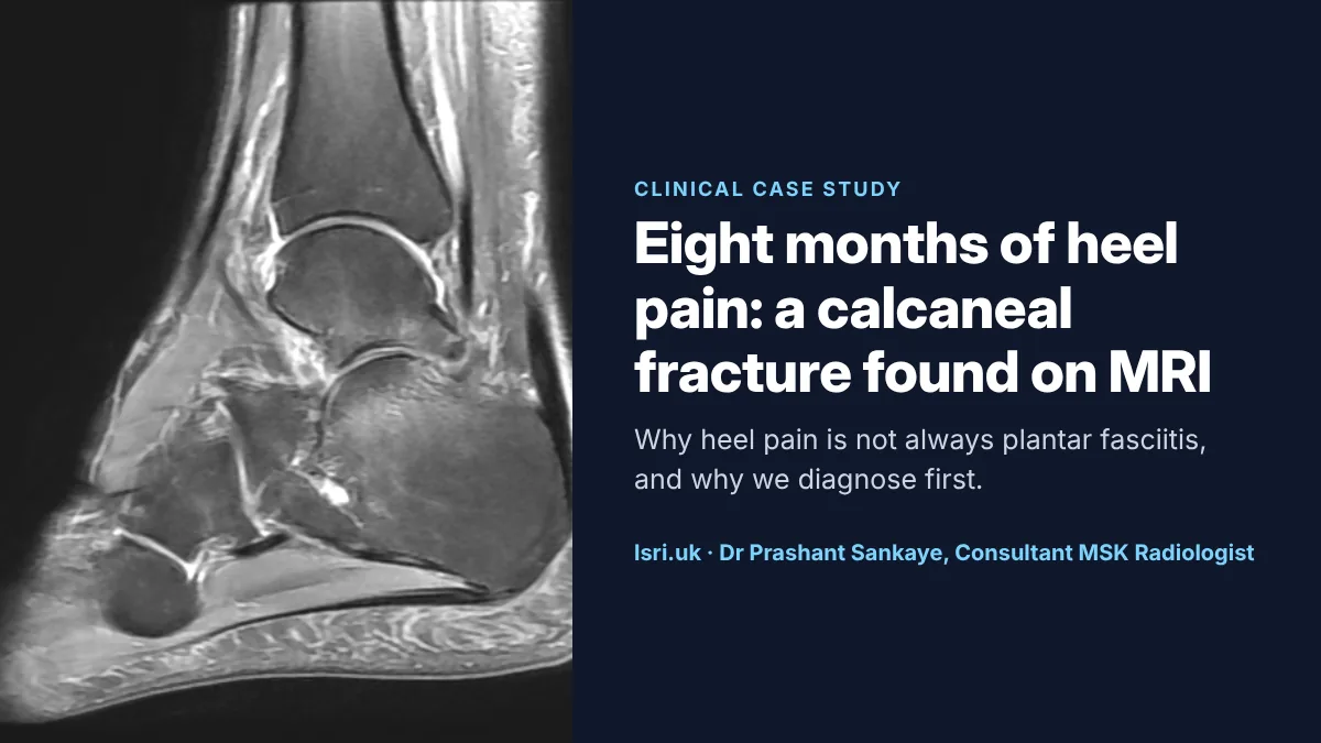
An adult patient developed heel pain. For about eight months it was managed with physiotherapy, as a soft-tissue problem, but the pain continued. An MRI scan at LSRI showed a fracture of the calcaneus, the large heel bone, with abnormal signal in the surrounding bone marrow.
Exercises for the plantar fascia, however well done, cannot heal a broken bone, and loading a fracture can keep it painful. Once the diagnosis was known, treatment could be aimed at the fracture.
This case is not about blame. Heel pain is common, plantar fasciitis is by far its most likely cause, and a heel bone fracture can be invisible on an X-ray. The lesson is about the pathway: when pain does not follow the expected course, the diagnosis needs to be reviewed, and that sometimes means a scan.
What the MRI showed
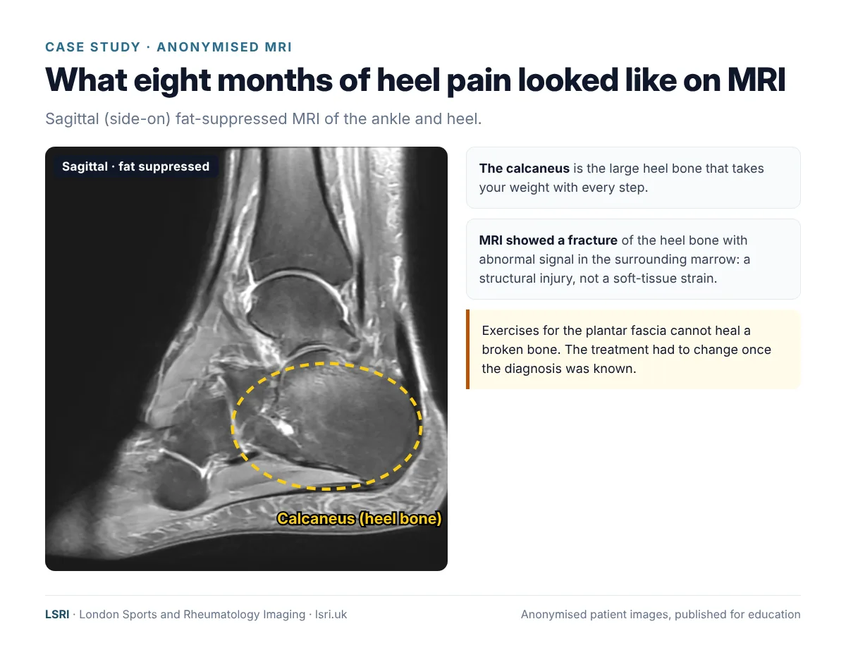
This image uses a fat-suppressed sequence, which darkens fat so that fluid stands out. Healthy heel bone marrow is mostly fat and looks dark on this sequence. Around a fracture, the marrow fills with fluid and turns brighter, and a fracture line may be visible as a thin line running through the bone. MRI shows these changes even when an X-ray looks normal.
How calcaneal fractures get missed
The calcaneus is the largest bone in the foot and takes the full weight of the body with every step. Fractures here are missed for two main reasons.
1. Stress fractures that X-rays cannot see
Stress injuries of the calcaneus are fairly common overuse injuries in military recruits and athletes,6 including long-distance runners.8 They often follow an increase in walking, running or standing, or a change to a harder surface.1 As with stress fractures elsewhere, X-rays are often normal in the first weeks, and some never show a fracture at all.2,5,6 In a study of military recruits with exercise-related heel or ankle pain, only 15% of the calcaneal stress injuries found on MRI were visible on X-ray.6 The good news is that the calcaneus is a “low-risk” stress fracture site: once diagnosed, these fractures usually heal well with a period of reduced loading.9
2. Subtle fractures that are easily overlooked
Some fractures of the heel bone are small or in awkward places. Fractures of the front part of the calcaneus (the anterior process), for example, can follow a twisting injury and are commonly mistaken for an ankle sprain.4 They can also be hard to see on X-ray: in one MRI study, only three of eight were visible on the X-rays.3 More generally, missed fractures are one of the commonest diagnostic errors in emergency care: in one UK study of an accident and emergency department, nearly 80% of diagnostic errors were missed fractures.10 A study of missed fractures in emergency radiology found that fractures of the foot were missed more often than those anywhere else, usually because they were subtle.11 None of this means anyone was careless. It reflects how difficult some fractures are to see on X-ray, and why MRI is valuable when pain does not settle.
Plantar fasciitis or a fracture?
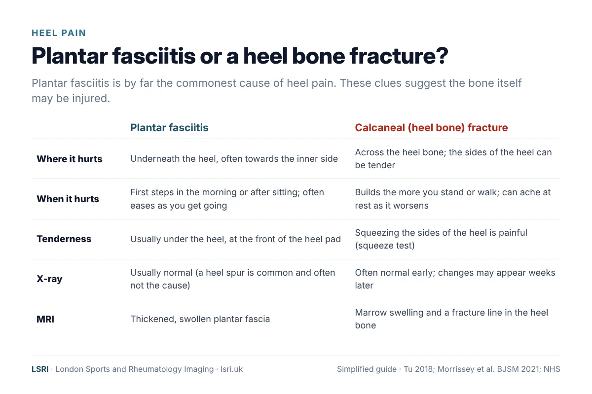
Plantar fasciitis typically causes pain under the heel, often towards the inner side, that is worst with the first steps in the morning or after sitting, and often eases as you get going.1,12 A calcaneal stress fracture tends to cause pain that comes on with activity and can later be present at rest, and pain when the sides of the heel are squeezed suggests the diagnosis.1 That said, the two can overlap, and examination alone cannot always tell them apart.
Plantar fasciitis itself is very treatable. A best-practice guide for plantar heel pain recommends taping, stretching and individualised education as the core treatment, with shockwave therapy and then custom insoles (orthoses) for people who do not improve enough.13 The point is not that physiotherapy is wrong. It is that physiotherapy for plantar fasciitis is the right treatment for plantar fasciitis, and the wrong treatment for a fracture.
Our approach: diagnose first, treat second
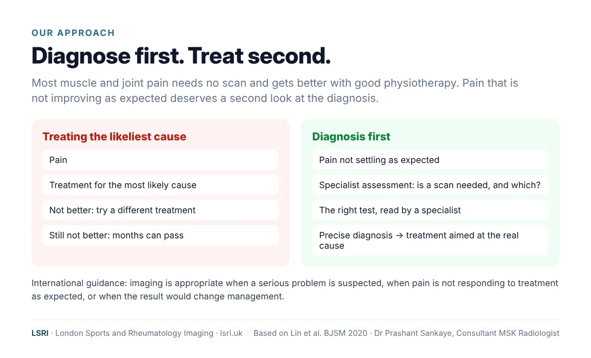
At LSRI, the principle that guides our work is simple: in musculoskeletal medicine, an accurate diagnosis comes first. Physiotherapy, injections and surgery are all powerful treatments, but each one works best when everyone knows exactly what is being treated.
“Our ethos is simple: diagnose first, diagnose accurately, and treat next. Physiotherapy is excellent treatment, but it works best when we know exactly what we are treating. When pain is not settling as expected, the most useful thing we can do is find out why.”
Dr Prashant Sankaye, Consultant MSK Radiologist, LSRI
This is not an argument for scanning everyone. International guidelines on musculoskeletal pain agree that routine imaging is not needed for most people, and that imaging should be used when a serious condition is suspected, when pain is not responding to treatment as expected, or when the result is likely to change management.7 Diagnosing first means using the right test at the right time, not the most tests.
What it does mean is not waiting months when the pattern is telling us something is wrong. Research on wrist injuries supports this. In a London trial of people attending A&E with a suspected scaphoid (wrist) fracture and a normal X-ray, offering an MRI straight away gave a diagnosis much sooner (on average in under 2 days rather than about 10), meant around half of those scanned needed no further clinic visits, found more fractures, and was cost-neutral at three months and cheaper by six months.14 That trial looked only at the wrist, but we believe the same principle applies elsewhere: when pain is not following the expected course, an early, accurate diagnosis can save time and months of uncertainty.
Why we share these cases
Every case in this series, from a hip stress fracture and a sacral insufficiency fracture to shoulder pain that needed an MRI and back pain that turned out to be cancer, carries the same lesson. LSRI is led by Dr Prashant Sankaye, a Consultant Musculoskeletal Radiologist with more than 20 years’ experience. Sharing these anonymised cases is how we try to help patients, physiotherapists, GPs and other clinicians recognise the patterns earlier, so that fewer people wait months for a diagnosis. We see that as part of our duty as clinicians.
What should you do if your heel pain isn’t settling?
- Give treatment a fair trial, and keep track. Plantar fasciitis can take months to settle, but it should be moving in the right direction.
- Notice the pattern. Pain that builds with every step, aches at rest, or hurts when the sides of the heel are squeezed may be coming from the bone.
- Think about what started it. A fall from a height, a twist, a jump in walking or running, or new hard surfaces are all clues.
- Ask for the diagnosis to be reviewed. It is reasonable to ask: “Could this be a fracture?”
- Ask about MRI if the cause is still unclear. A normal X-ray does not rule out a stress fracture.
- Know when it is urgent. If you have severe heel pain after an injury, cannot walk, heard a snap, grinding or popping noise at the time of the injury, or your foot or ankle has changed shape, get urgent advice from NHS 111 (by phone or online).12
How LSRI can help
- Advice before you book. Call us to discuss whether a scan is likely to help, and which one.
- Specialist reporting. Your MRI is reported by a consultant musculoskeletal radiologist within 24 hours.
- Clear pricing. MRI from £375 per body part, including the scan, a specialist report and access to your images.
- Time to talk. MRI with a consultation, to go through the results and next steps, is from £425.
See our private MRI scans page, our page on plantar fasciitis, or our guide on stress fractures that don’t show on X-ray.
Frequently asked questions
Can you walk on a fractured heel?
Sometimes, yes, which is one reason heel fractures are missed. Stress fractures and small fractures may allow walking, although it is usually painful. Walking on an undiagnosed fracture can delay healing, so if heel pain is not settling, get it checked.
Will a calcaneal stress fracture show on an X-ray?
Often not in the early weeks. Changes may appear later as the bone heals, but some stress fractures never show on X-ray. MRI is the most accurate test.
How do I know if my heel pain is plantar fasciitis or a fracture?
Plantar fasciitis is usually worst with the first steps in the morning and eases as you move. Fracture pain tends to build with walking, and squeezing the sides of the heel hurts. But the symptoms can overlap, so if your pain is not improving as expected, ask for the diagnosis to be reviewed.
How long does a calcaneal stress fracture take to heal?
Most heal in roughly 8 to 12 weeks with reduced loading, sometimes using a walking boot,8 followed by a gradual return to activity. Larger or displaced fractures from a significant injury are managed differently and may need an orthopaedic opinion.
Do I need a GP referral for a private foot MRI?
No GP referral is needed in most cases if you are paying for yourself. If you are claiming through private medical insurance, your insurer may require a referral, so check with them first. We always recommend sharing your results with your GP.
Book an appointment
Call 0203 633 5040 or book online. Clinics at Harley Street (W1G 9QJ) and Harrow (HA2 7SJ). If you have emergency symptoms, go to A&E or call 999 instead.
References
- Tu P. Heel pain: diagnosis and management. American Family Physician 2018;97(2):86–93. aafp.org
- Tins BJ, Garton M, Cassar-Pullicino VN, et al. Stress fracture of the pelvis and lower limbs including atypical femoral fractures: a review. Insights into Imaging 2015;6(1):97–110. doi:10.1007/s13244-014-0371-z
- Ouellette H, Salamipour H, Thomas BJ, et al. Incidence and MR imaging features of fractures of the anterior process of calcaneus in a consecutive patient population with ankle and foot symptoms. Skeletal Radiology 2006;35(11):833–837. doi:10.1007/s00256-006-0154-9
- Judd DB, Kim DH. Foot fractures frequently misdiagnosed as ankle sprains. American Family Physician 2002;66(5):785–794. aafp.org
- Wright AA, Hegedus EJ, Lenchik L, et al. Diagnostic accuracy of various imaging modalities for suspected lower extremity stress fractures: a systematic review with evidence-based recommendations for clinical practice. American Journal of Sports Medicine 2016;44(1):255–263. doi:10.1177/0363546515574066
- Sormaala MJ, Niva MH, Kiuru MJ, et al. Stress injuries of the calcaneus detected with magnetic resonance imaging in military recruits. Journal of Bone and Joint Surgery (American) 2006;88(10):2237–2242. doi:10.2106/JBJS.E.01447. PubMed
- Lin I, Wiles L, Waller R, et al. What does best practice care for musculoskeletal pain look like? Eleven consistent recommendations from high-quality clinical practice guidelines: systematic review. British Journal of Sports Medicine 2020;54(2):79–86. doi:10.1136/bjsports-2018-099878
- Robertson GAJ, Wood AM. Lower limb stress fractures in sport: optimising their management and outcome. World Journal of Orthopedics 2017;8(3):242–255. doi:10.5312/wjo.v8.i3.242
- Kaeding CC, Yu JR, Wright R, et al. Management and return to play of stress fractures. Clinical Journal of Sport Medicine 2005;15(6):442–447. doi:10.1097/01.jsm.0000188207.62608.35
- Guly HR. Diagnostic errors in an accident and emergency department. Emergency Medicine Journal 2001;18(4):263–269. doi:10.1136/emj.18.4.263
- Wei CJ, Tsai WC, Tiu CM, et al. Systematic analysis of missed extremity fractures in emergency radiology. Acta Radiologica 2006;47(7):710–717. doi:10.1080/02841850600806340
- NHS. Heel pain. nhs.uk
- Morrissey D, Cotchett M, Said J’Bari A, et al. Management of plantar heel pain: a best practice guide informed by a systematic review, expert clinical reasoning and patient values. British Journal of Sports Medicine 2021;55(19):1106–1118. doi:10.1136/bjsports-2019-101970
- Rua T, Malhotra B, Vijayanathan S, et al. Clinical and cost implications of using immediate MRI in the management of patients with a suspected scaphoid fracture and negative radiographs: results from the SMaRT trial. Bone & Joint Journal 2019;101-B(8):984–994. doi:10.1302/0301-620X.101B8.BJJ-2018-1590.R1
About this case study
Patient privacy: the image is anonymised, with no names, dates or other identifying details, and non-essential details of the case have been left out. It is shared for education. Purpose: written by the LSRI clinical team to help the public recognise when heel pain needs a closer look, based on the research and guidance listed above. It is general information, not a diagnosis; if you are worried about your own symptoms, speak to your GP. The case is not a criticism of any clinician: physiotherapy is the usual and appropriate first treatment for most heel pain.