Written by: LSRI Editorial Team
Medically reviewed by Dr Prashant Sankaye, Consultant MSK Radiologist ·
Last Updated: October 14, 2026
Read Time: 11 Minutes
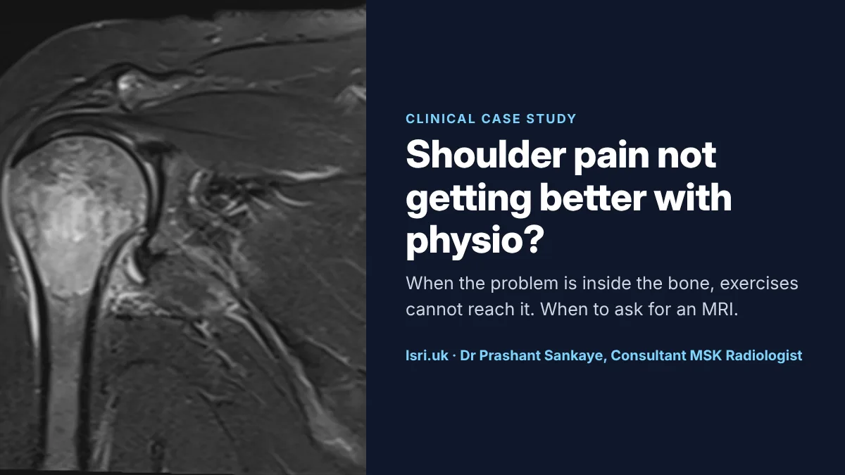
Shoulder pain is one of the most common reasons people see a physiotherapist, and for good reason: most shoulder pain comes from the rotator cuff tendons and the surrounding tissues, and a structured exercise programme helps the majority of people. But physiotherapy works best when the diagnosis is right. When shoulder pain does not improve as expected, the answer is not always “more of the same”. Sometimes the problem is somewhere exercises cannot reach, such as inside the bone itself. This anonymised case from our clinic, in which months of physiotherapy for shoulder pain were followed by an MRI showing an abnormality inside the bone, explains when shoulder pain needs a scan and which scan answers which question.
Key points
- Most shoulder pain is related to the rotator cuff and improves with physiotherapy. A scan is not needed for everyone.1,2
- UK shoulder pathways recommend reviewing the diagnosis, and considering referral, when pain is not improving after a good course of treatment, usually within around three months.2
- Ultrasound is excellent for tendons, but it cannot see inside bone. A normal ultrasound does not rule out problems such as arthritis, bone lesions or a tumour.1,3
- MRI shows the tendons, cartilage, labrum and the bone marrow in one scan.
- Warning signs such as a lump or swelling, fever or feeling unwell, a hot or red shoulder, or sudden weakness after an injury need prompt assessment.1,2,4
The case: months of physiotherapy, then an MRI
An adult patient had shoulder pain that was treated with physiotherapy for several months. That was a reasonable approach: it is how most shoulder pain is managed, and most people improve with it. But this pain did not settle.
When an MRI scan was done at LSRI, it showed that the main problem was not in the tendons at all. There was an extensive abnormality inside the bone of the humeral head, the ball of the shoulder joint. The bone marrow there had lost its normal fatty signal and was bright on fluid-sensitive images. Further assessment showed that the cause was a tumour in the bone; to protect the patient’s privacy, we have not given further details. That is not something physiotherapy can treat, and the diagnosis changed the next steps entirely.
This case is not about blame. Shoulder pain from a problem inside the bone can feel very like rotator cuff pain, and the first tests used for shoulder pain, such as ultrasound, are not designed to see inside bone. The lesson is about timing: when pain does not follow the expected course, the diagnosis needs to be reviewed, and that sometimes means an MRI.
What the MRI showed
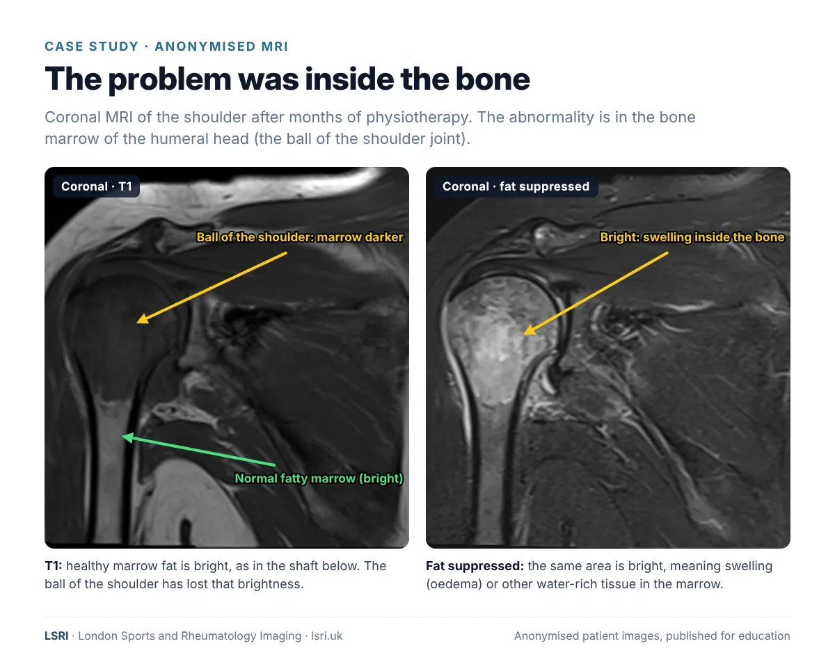
In adults, the bone marrow of the upper arm bone is mostly fat, and fat looks bright on a T1-weighted image. You can see that brightness in the shaft of the bone lower down. In the ball of the shoulder, the marrow has lost that brightness: something has replaced or displaced the fat. On the fat-suppressed image, which darkens fat so that water stands out, the same area is bright. That combination tells the radiologist that there is swelling (bone marrow oedema) or other abnormal, water-rich tissue inside the bone.
Reading the two sequences together, along with the other views, the pattern of the change and the patient’s history, is how a specialist radiologist narrows down the cause. Some normal variations, such as patches of blood-forming red marrow in the shoulder, can look similar, which is why experience in reading musculoskeletal MRI matters.5
What can cause problems inside the bone of the shoulder?
Bright marrow on a fluid-sensitive MRI is a sign, not a diagnosis. Possible causes, in the shoulder as elsewhere in the skeleton, include:6,7
- Bone bruising or a hidden (occult) fracture after an injury or fall, which may not show on X-ray.
- Avascular necrosis (osteonecrosis), where part of the bone loses its blood supply. Our case study on four years of shoulder pain and avascular necrosis explains this condition.8
- Arthritis, with bone changes under worn cartilage.
- Infection in the bone or joint, which is uncommon but urgent.
- Inflammatory conditions, such as inflammatory arthritis.
- Bone tumours. Many bone lesions are benign, but cancer can also spread to bone from elsewhere (metastasis), and the upper arm bone is the second most common long bone for this to affect.9
Some of these are minor and settle on their own; some need urgent treatment. What they have in common is that they cannot be diagnosed by examination alone, and many need treatment beyond, or instead of, an exercise programme.
Why most shoulder pain still starts with physiotherapy
It is important to say clearly: for most people, physiotherapy is the right first treatment for shoulder pain. The British Elbow and Shoulder Society (BESS) and British Orthopaedic Association pathways for the most common type of shoulder pain, called subacromial pain, recommend exercise-based treatment first, with imaging reserved for when red flags are present or symptoms are not improving.1,2 Scanning everyone early would not help most people, and MRI often shows age-related changes in the tendons of people with no pain at all.
The question is not whether physiotherapy is a good idea. It is what to do when it is not working.
When should shoulder pain be scanned?
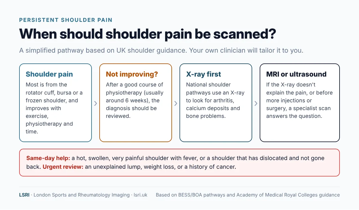
Based on UK shoulder pathways, the main situations are:1,2
- Red flags (see below), at any stage.
- Pain that has not improved after a good-quality course of physiotherapy, usually within around 6 to 12 weeks. The 2025 BESS pathway recommends referral if symptoms have not improved by about 12 weeks, and an X-ray if there are red flags or pain has lasted more than about four weeks.2
- Loss of strength or movement after an injury, which may mean a tendon tear that is best repaired early.
- When the result would change treatment, for example before an injection or surgery.
Shoulder red flags
Seek prompt medical assessment if shoulder pain comes with any of these:1,2,4
- a history of cancer, or a new lump or swelling;
- unexplained weight loss, fever, or feeling generally unwell;
- a hot, red or swollen joint;
- severe, constant pain, especially at night, that does not ease with rest;
- sudden weakness or inability to lift the arm after an injury, or a dislocation;
- pins and needles that do not go away, or numbness in the arm or shoulder.
If you have chest pain, breathlessness or pain in the left shoulder or arm that comes on suddenly, call 999: shoulder pain can occasionally come from the heart.
Which scan answers which question?
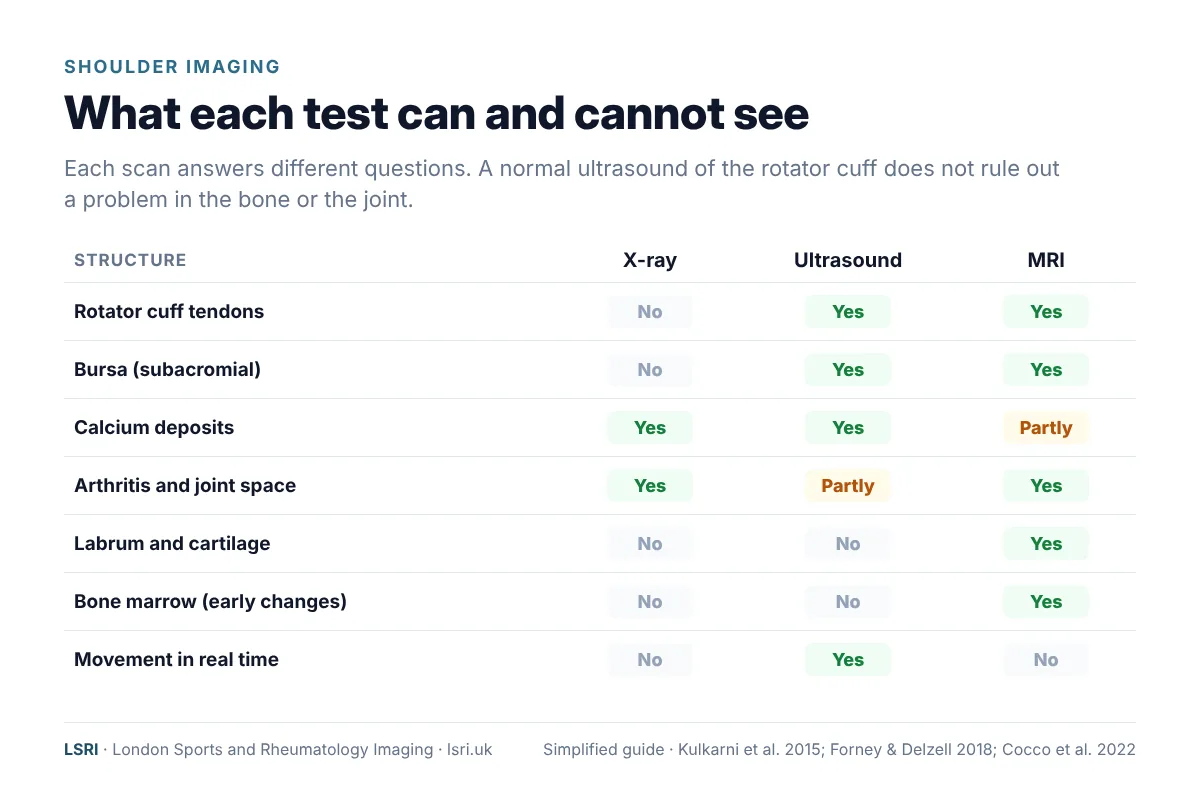
- X-ray is quick and shows the bones, the joint space and calcium deposits. It is often the first test when pain has persisted or there are red flags. It does not show tendons, and early changes inside the bone may not be visible.
- Ultrasound is excellent for the rotator cuff tendons and the bursa, and it can watch the tendons move in real time. It is also used to guide injections. But sound waves do not pass through bone, so ultrasound cannot assess the bone marrow.3,10 The BESS/BOA pathway notes that a normal rotator cuff ultrasound does not exclude serious problems such as a tumour or osteoarthritis.1
- MRI shows the tendons, muscles, cartilage, labrum and joint lining, and it is the only one of the three that shows the bone marrow directly. That is why it found the problem in this case.
For more detail, see our guides to what a shoulder MRI shows and shoulder ultrasound scans, or read MRI or ultrasound for joint pain.
What should you do if your shoulder pain isn’t getting better?
- Give treatment a fair trial, and keep track. Note whether pain, sleep and movement are improving week by week.
- If it is not improving after several weeks, say so. Ask your physiotherapist or GP whether the diagnosis should be reviewed.
- Ask what has been ruled out. If you have had an ultrasound, remember that it does not look inside the bone.
- Ask about MRI if the cause is still unclear. MRI is the test that shows the bone marrow as well as the soft tissues.
- Do not ignore red flags. Severe night pain that does not ease with rest, a lump, a history of cancer or feeling unwell need prompt assessment.
How LSRI can help
- Advice before you book. Call us to discuss whether a scan is likely to help, and which one: X-ray, ultrasound or MRI.
- Specialist reporting. Your MRI is reported by a consultant musculoskeletal radiologist within 24 hours. If the scan shows something serious, we contact you and your GP so that urgent next steps are not delayed.
- Clear pricing. MRI from £375 per body part, including the scan, a specialist report and access to your images.
- Time to talk. MRI with a consultation, to go through the results and next steps, is from £425.
See our private MRI scans page, ask for a second opinion on a scan you have already had, or read why we diagnose first and treat second.
Frequently asked questions
How long should I do physio for shoulder pain before getting a scan?
There is no single rule, but UK pathways suggest reviewing the diagnosis if pain is not improving after around 6 to 12 weeks of good-quality treatment, and sooner if there are red flags. If your pain is getting worse rather than better, do not wait.
My shoulder ultrasound was normal. Why does it still hurt?
Ultrasound is excellent for tendons and bursae, but it cannot see inside the bone or assess the labrum and most of the joint cartilage. A normal ultrasound rules out some causes but not all. If pain persists, an MRI may be the next step.
What does bone marrow oedema in the shoulder mean?
It means there is extra fluid inside the bone, usually as a reaction to something: an injury, a stress on the bone, arthritis, loss of blood supply, inflammation, infection, or occasionally a tumour. It is a sign that needs interpreting in context, not a diagnosis on its own. Your radiologist and doctor will explain what it means for you.
Is shoulder pain at night serious?
Night pain is common with rotator cuff problems and frozen shoulder, and on its own it is usually not serious. But severe, constant night pain that does not ease with rest or change of position, especially with other warning signs, should be checked promptly.
Do I need a GP referral for a private shoulder MRI?
No GP referral is needed in most cases if you are paying for yourself. If you are claiming through private medical insurance, your insurer may require a referral, so check with them first. We always recommend sharing your results with your GP.
Book an appointment
Call 0203 633 5040 or book online. Clinics at Harley Street (W1G 9QJ) and Harrow (HA2 7SJ). If you have emergency symptoms, go to A&E or call 999 instead.
References
- Kulkarni R, Gibson J, Brownson P, et al. BESS/BOA patient care pathways: subacromial shoulder pain. Shoulder & Elbow 2015;7(2):135–143. doi:10.1177/1758573215576456
- Pandey RA, Singh HP, on behalf of the BESS Subacromial Pain Working Group. BESS patient care pathway: subacromial pain. Shoulder & Elbow 2025;17(6):713–724. doi:10.1177/17585732251374282
- Forney MC, Delzell PB. Musculoskeletal ultrasonography basics. Cleveland Clinic Journal of Medicine 2018;85(4):283–300. doi:10.3949/ccjm.85a.17014
- NHS. Shoulder pain. nhs.uk
- Richardson ML, Patten RM. Age-related changes in marrow distribution in the shoulder: MR imaging findings. Radiology 1994;192(1):209–215. doi:10.1148/radiology.192.1.8208940
- Starr AM, Wessely MA, Albastaki U, et al. Bone marrow edema: pathophysiology, differential diagnosis, and imaging. Acta Radiologica 2008;49(7):771–786. doi:10.1080/02841850802161023
- Hayes CW, Conway WF, Daniel WW. MR imaging of bone marrow edema pattern: transient osteoporosis, transient bone marrow edema syndrome, or osteonecrosis. RadioGraphics 1993;13(5):1001–1011. doi:10.1148/radiographics.13.5.8210586
- Castillo Mercado JS, Rojas Liévano J, Zaldivar B, et al. Atraumatic osteonecrosis of the humeral head: pathophysiology and current concepts of evaluation and treatment. JSES Reviews, Reports, and Techniques 2022;2(3):277–284. doi:10.1016/j.xrrt.2022.02.005
- Scotti C, Camnasio F, Peretti GM, et al. Modular prostheses in the treatment of proximal humerus metastases: review of 40 cases. Journal of Orthopaedics and Traumatology 2008;9(1):5–10. doi:10.1007/s10195-008-0097-0
- Cocco G, Ricci V, Villani M, et al. Ultrasound imaging of bone fractures. Insights into Imaging 2022;13(1):189. doi:10.1186/s13244-022-01335-z
About this case study
Patient privacy: the images are anonymised, with no names, dates or other identifying details, and non-essential details of the case have been left out. They are shared for education. Purpose: written by the LSRI clinical team to help the public understand when shoulder pain needs a closer look, based on the research and guidance listed above. It is general information, not a diagnosis; if you are worried about your own symptoms, speak to your GP. The case is not a criticism of any clinician: physiotherapy is the usual and appropriate first treatment for most shoulder pain.