Written by: LSRI Editorial Team
Medically reviewed by Dr Prashant Sankaye, Consultant MSK Radiologist ·
Last Updated: October 14, 2026
Read Time: 11 Minutes
Written by the LSRI clinical team and medically reviewed by Dr Prashant Sankaye, Consultant MSK Radiologist, on 2 October 2026.
If you have joint pain, you may have been told you need “a scan”, without being told which one. MRI and ultrasound are both excellent tests, but they are good at different things. Choosing the right one first can save time, avoid a second appointment and get you to the right treatment sooner. This guide explains what each scan shows best, where an X-ray fits in, and when you may not need a scan at all. For details of our services, see our ultrasound scans and MRI scans pages.
MRI or ultrasound for joint pain: the short answer
- Ultrasound is usually the best first test for problems close to the surface: tendons, bursae, many ligaments, muscles, soft-tissue lumps and fluid around a joint. It is quick, uses no radiation, and can look at the area while it moves. It is also the scan used to guide injections.
- MRI is usually the best test for problems deep inside a joint or inside bone: cartilage, the menisci and cruciate ligaments in the knee, the labrum in the hip and shoulder, bone bruises, stress fractures and the spine.
- X-ray is a quick way to look at bones, joint space and alignment, for example after a fall or when arthritis is suspected.
- Sometimes no scan is needed. A careful history and examination are often enough to make the diagnosis and start treatment.
What ultrasound shows best
Ultrasound uses high-frequency sound waves to build a live picture of the tissues under the probe. The NHS notes that ultrasound scans do not use any radiation, unlike X-rays or CT scans, and that most scans take around 10 to 45 minutes1.
For joint and muscle problems, ultrasound is particularly good at showing:
- Tendons: for example the rotator cuff in the shoulder, the Achilles tendon, the tendons on the outside of the hip, and the tendons involved in tennis and golfer’s elbow.
- Bursae and fluid: a swollen bursa, a joint effusion or a Baker’s cyst behind the knee.
- Ligaments and muscles near the surface: for example ankle ligament injuries and many muscle tears.
- Soft-tissue lumps: ultrasound is usually the first test for a lump under the skin. See our lumps and bumps clinic.
- Inflammation of the joint lining: in suspected inflammatory arthritis, ultrasound can show inflamed joint lining (synovitis) and tendon sheaths. European rheumatology recommendations note that ultrasound and MRI can detect joint inflammation more reliably than clinical examination alone, and can help when the diagnosis is in doubt2.
Two further strengths are unique to ultrasound. It is dynamic: we can move your joint during the scan to see a tendon catching, snapping or pinching. And the other side can be scanned in the same appointment for comparison. Because the picture is live, the radiologist can often explain what they see while you are still in the room.
Limits: sound waves do not pass through bone, so ultrasound cannot see inside bones or deep into the middle of some joints. It cannot assess the spine. The quality of an ultrasound scan also depends heavily on the skill and experience of the person doing it.
What MRI shows best
MRI uses a strong magnetic field and radio waves to produce detailed pictures in several planes. It gives a complete view of a joint, including the structures ultrasound cannot reach:
- Inside the knee: the menisci, the cruciate ligaments and the joint cartilage. Our knee MRI patient guide explains more.
- Inside the hip and shoulder: the labrum and cartilage, and the whole rotator cuff. See what a shoulder MRI shows.
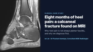
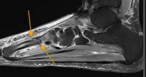
- Bone: bone bruises, stress fractures (which are often invisible on an early X-ray) and changes in the bone marrow.
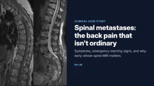
- The spine and nerves: discs, nerve roots and the spinal canal. See spine MRI for back pain.
Limits: an MRI takes longer than an ultrasound (the NHS says between 15 minutes and an hour3), you need to lie still, and the scanner is noisy. The images are taken with the joint still, so problems that only appear with movement can be missed. Some people cannot have an MRI, or need extra checks first, for example if they have a pacemaker or certain other metal implants, and you should say if you are or might be pregnant. If you feel anxious in enclosed spaces, tell us when you book. Our guide to MRI scans covers what to expect.
MRI is also very sensitive. It often shows age-related changes, such as wear in a meniscus or disc, that are common in people with no pain at all4,5. That is why the scan should always be interpreted alongside your symptoms and examination, ideally by a specialist musculoskeletal radiologist.
Is ultrasound as good as MRI?
For some questions, yes. A Cochrane review of studies in people with shoulder pain found that MRI, MR arthrography and ultrasound performed similarly for detecting full-thickness rotator cuff tears. All were less reliable for partial-thickness tears, and ultrasound more so than MRI. In other situations, such as a suspected meniscal tear or a stress fracture, MRI gives information ultrasound simply cannot. The honest answer is that the “better” scan depends on the question being asked.
Where X-ray fits in
An X-ray is quick and widely available. It shows bone, the space between the bones in a joint (which narrows in osteoarthritis), alignment, and calcium deposits such as those in calcific tendinitis. It uses a small dose of radiation7. X-rays show little of the soft tissues, so a normal X-ray does not rule out a tendon, ligament or cartilage injury. See our X-ray page.
When you may not need a scan at all
A scan is only useful if the result could change what happens next. Two examples:
- Osteoarthritis: adults aged 45 or over can be diagnosed with osteoarthritis clinically, without imaging, if they have activity-related joint pain and any morning stiffness lasts no longer than 30 minutes. Imaging findings often match symptoms poorly. A scan may still help if the symptoms are unusual, if something else is suspected, or before an injection or surgery is planned.
- Low back pain: routine imaging is not usually needed for low back pain, with or without sciatica, outside specialist care. In specialist settings, a scan is used when the result is likely to change management.
If a scan will not change your treatment, we will say so.
Common problems and the usual first scan
This table is a general guide, not a diagnosis. The right test for you depends on your history and examination.
| Symptom or suspected problem | Usual first scan | When MRI (or another test) may be needed |
|---|---|---|
| Shoulder pain when lifting the arm (rotator cuff, bursitis) | Ultrasound | Suspected labral tear, instability, or planning surgery |
| Knee injury with twisting, locking or giving way | MRI | Ultrasound can check for fluid or a Baker’s cyst |
| Gradual knee pain in someone aged 45 or over | Often no scan; X-ray if unclear | MRI if symptoms are unusual or not settling |
| Pain on the outside of the hip, worse lying on that side | Ultrasound | MRI if the diagnosis is unclear |
| Deep groin pain, catching or clicking in the hip | MRI | X-ray to look at the shape of the joint |
| Tennis or golfer’s elbow | Ultrasound | MRI occasionally, if pain persists or the diagnosis is unclear |
| Achilles or heel pain (including plantar fasciitis) | Ultrasound | MRI if a bone problem such as a stress fracture is suspected |
| Suspected stress fracture | MRI | An early X-ray is often normal |
| Swollen, stiff small joints with long morning stiffness | Ultrasound, alongside blood tests and a rheumatology assessment | MRI in selected cases |
| A lump under the skin | Ultrasound | MRI if the lump is large, deep or the ultrasound is not conclusive |
| Back pain with leg pain (sciatica) | Often no scan at first | MRI if the result would change treatment, or for red-flag symptoms |
When to get urgent help instead
Do not wait for a routine scan if you have:
- a hot, red, swollen joint with a fever or feeling unwell;
- an injury after which you cannot put weight on the leg, or there is an obvious deformity;
- back pain with numbness around your genitals or bottom, new problems passing urine or controlling your bowels, or weakness in both legs.
Go to A&E or call 999 in these situations. For other urgent concerns, contact your GP or NHS 111.
How we choose the right scan at LSRI
At LSRI, scans are led by Dr Prashant Sankaye, a Consultant MSK Radiologist. Because we offer ultrasound, MRI and X-ray, we can recommend the test that answers your question rather than the one that happens to be available, using national guidance such as the Royal College of Radiologists’ iRefer11.
- If an ultrasound scan answers the question, you may not need an MRI at all. If it shows that an injection is appropriate, the same radiologist can guide it precisely. See our ultrasound-guided injections.
- If you need an MRI, we can arrange it at Harley Street; for Harrow patients it is arranged at a partner centre. If you live further away, we can arrange it at one of 70+ UK locations including partner centres.
- Your MRI is reported by a consultant musculoskeletal radiologist within 24 hours.
- Private MRI from £375 per body part, reported by a consultant radiologist within 24 hours.
- Already had a scan elsewhere? We also offer second opinions on existing images and reports.
Frequently asked questions
Is ultrasound or MRI safer?
Neither uses ionising radiation. Diagnostic ultrasound is considered very safe. MRI is very safe for most people, but the strong magnet means some implants and devices need to be checked first, so we will ask you a set of safety questions before your scan.
Can I choose which scan I have?
Yes, you can tell us your preference. We will explain which scan is most likely to answer your question, and why, so that you can decide together with us.
Do I need a GP referral?
No GP referral is needed in most cases if you are paying for yourself. If you are claiming through private medical insurance, your insurer may require a referral from a GP or specialist, so check with them first.
When will I get my results?
With ultrasound, the radiologist can often explain the findings during the scan, and a written report follows. MRI scans are usually reported within 24 hours.
What if my MRI shows “wear and tear”?
Age-related changes are very common on MRI, including in people without pain. What matters is whether the findings fit your symptoms. Our guide to understanding your MRI results explains common terms.
Book an appointment
Call 0203 633 5040 or book online. If you are not sure which scan you need, call us and we will help you choose. Clinics at Harley Street (W1G 9QJ) and Harrow (HA2 7SJ).
References
- NHS. Ultrasound scan. https://www.nhs.uk/tests-and-treatments/ultrasound-scan/ (page last reviewed 25 February 2025)
- Colebatch AN, Edwards CJ, Østergaard M, et al. EULAR recommendations for the use of imaging of the joints in the clinical management of rheumatoid arthritis. Ann Rheum Dis 2013;72(6):804-814. doi:10.1136/annrheumdis-2012-203158
- NHS. MRI scan. https://www.nhs.uk/tests-and-treatments/mri-scan/ (page last reviewed 11 September 2025)
- Brinjikji W, Luetmer PH, Comstock B, et al. Systematic literature review of imaging features of spinal degeneration in asymptomatic populations. AJNR Am J Neuroradiol 2015;36(4):811-816. doi:10.3174/ajnr.A4173
- Culvenor AG, Øiestad BE, Hart HF, Stefanik JJ, Guermazi A, Crossley KM. Prevalence of knee osteoarthritis features on magnetic resonance imaging in asymptomatic uninjured adults: a systematic review and meta-analysis. Br J Sports Med 2019;53(20):1268-1278. doi:10.1136/bjsports-2018-099257
- Lenza M, Buchbinder R, Takwoingi Y, Johnston RV, Hanchard NCA, Faloppa F. Magnetic resonance imaging, magnetic resonance arthrography and ultrasonography for assessing rotator cuff tears in people with shoulder pain for whom surgery is being considered. Cochrane Database Syst Rev 2013;(9):CD009020. doi:10.1002/14651858.CD009020.pub2
- NHS. X-ray. https://www.nhs.uk/tests-and-treatments/x-ray/
- Sakellariou G, Conaghan PG, Zhang W, et al. EULAR recommendations for the use of imaging in the clinical management of peripheral joint osteoarthritis. Ann Rheum Dis 2017;76(9):1484-1494. doi:10.1136/annrheumdis-2016-210815
- Chou R, Qaseem A, Snow V, et al. Diagnosis and treatment of low back pain: a joint clinical practice guideline from the American College of Physicians and the American Pain Society. Ann Intern Med 2007;147(7):478-491. doi:10.7326/0003-4819-147-7-200710020-00006
- Lin I, Wiles L, Waller R, et al. What does best practice care for musculoskeletal pain look like? Eleven consistent recommendations from high-quality clinical practice guidelines: systematic review. Br J Sports Med 2020;54(2):79-86. doi:10.1136/bjsports-2018-099878
- The Royal College of Radiologists. iRefer: making the best use of clinical radiology. https://www.irefer.org.uk/