Written by: LSRI Editorial Team
Medically reviewed by Dr Prashant Sankaye, Consultant MSK Radiologist ·
Last Updated: October 14, 2026
Read Time: 12 Minutes
Written by the LSRI clinical team and medically reviewed by Dr Prashant Sankaye, Consultant MSK Radiologist, on 5 October 2026.
Most shoulder pain comes from the soft tissues, such as the rotator cuff, the bursa or a frozen shoulder, and gets better with exercise, physiotherapy and time. But when shoulder pain goes on for months or years despite treatment, the diagnosis deserves a second look. In this anonymised case from our clinic, four years of shoulder pain treated with physiotherapy and steroid injections turned out, on MRI, to be osteoarthritis of the shoulder joint together with avascular necrosis (osteonecrosis) of the humeral head, the ball of the shoulder. This guide explains what avascular necrosis of the shoulder is, how it progresses, why early diagnosis widens the treatment options, and when persistent shoulder pain should be scanned. For general information, see what a shoulder MRI shows.
Key points
- Avascular necrosis (AVN) means bone has died because its blood supply was interrupted. The humeral head is the second most common site after the hip.
- Corticosteroid medicines are the most common cause of AVN without an injury, mainly high-dose or long courses of tablets or drips. Other causes include alcohol, sickle cell disease, some cancer treatments and injury.
- Early AVN does not show on X-ray. MRI is the most sensitive test and can find it before the bone collapses.
- Once the ball of the joint collapses, arthritis follows and treatment options narrow, often to joint replacement.
- UK shoulder guidance uses an X-ray when shoulder pain is not improving, and recommends specialist review when the diagnosis is in doubt.
The case: four years of shoulder pain
An adult patient had shoulder pain for about four years. Over that time it was treated as a soft-tissue shoulder problem, with physiotherapy and steroid injections. The pain persisted.
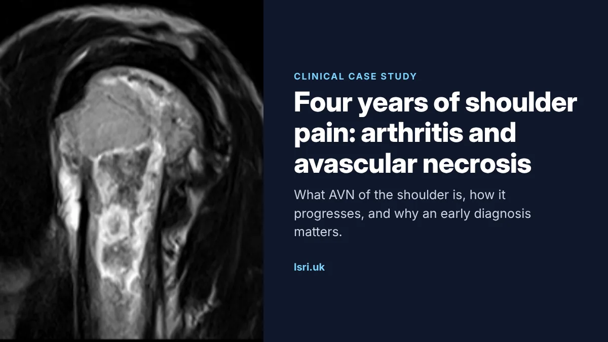
When the patient had an MRI scan at LSRI, it showed osteoarthritis of the shoulder joint and osteonecrosis of the humeral head, with the same process affecting the upper shaft of the humerus (the upper arm bone). These are structural problems in the bone and joint, which explains why treatments aimed at the soft tissues had not resolved the pain.
This is not a criticism of the clinicians involved. Shoulder pain is common, it is usually a soft-tissue problem, and physiotherapy is the right first treatment for most people. The lesson is that pain which persists despite good treatment is itself a reason to revisit the diagnosis.
What the MRI showed
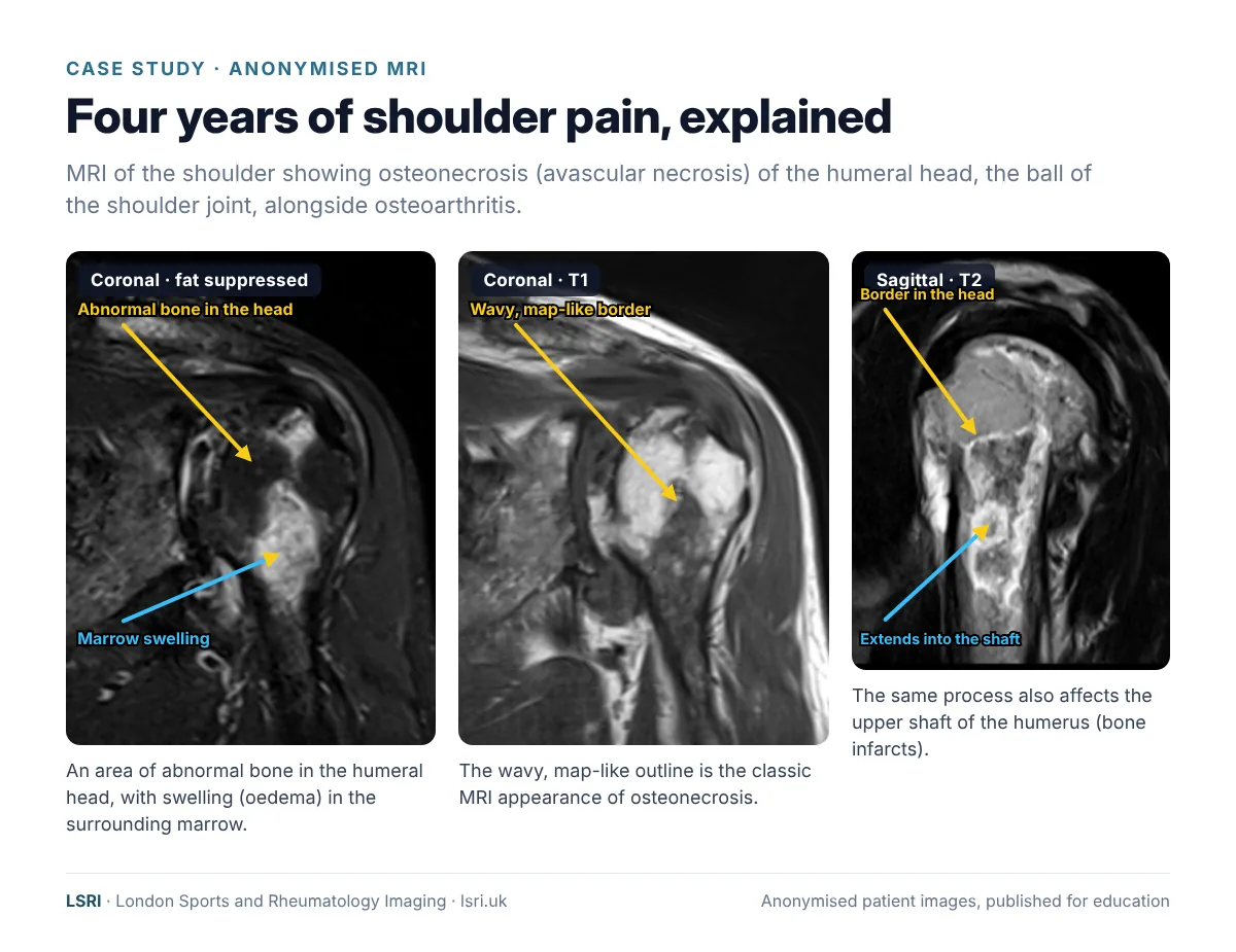
The key finding is the wavy, map-like (serpiginous) border in the humeral head. It marks the edge between dead bone and the living bone around it, which reacts by forming a rim of repair tissue. On T1-weighted images this border is dark; on fluid-sensitive images there is often a bright inner line alongside it, a pattern radiologists call the “double-line sign”. Swelling (oedema) in the surrounding marrow suggests the process is active. The sagittal image shows similar changes further down the shaft, called bone infarcts. Alongside this, the joint showed the wear of osteoarthritis.
What is avascular necrosis of the shoulder?
Bone is living tissue with its own blood supply. If that supply is interrupted, the affected bone cells die. This is called avascular necrosis or osteonecrosis. The dead bone gradually weakens, and the smooth, curved joint surface above it can crack and collapse.
The hip (femoral head) is the most common site, accounting for about 80% of cases, and the humeral head is the second, at about 10%.1,2 Recognised causes and risk factors include:3
- corticosteroid medicines, the most common cause of osteonecrosis not caused by injury, especially high doses or long courses;
- alcohol, in a dose-dependent way;
- sickle cell disease and other blood disorders;
- lupus (SLE) and some other autoimmune conditions;
- chemotherapy and radiotherapy;
- diving-related decompression illness, Gaucher disease, pancreatitis and pregnancy;
- injury, particularly complex fractures of the top of the humerus;2
- and sometimes no cause is found (idiopathic).
Osteonecrosis without an injury often affects more than one joint. Atraumatic osteonecrosis of the humeral head is seldom isolated and frequently occurs alongside osteonecrosis elsewhere, including the hips.4 When it is found, a specialist will usually look for an underlying cause and may recommend imaging of other joints.5
Can steroid injections cause avascular necrosis?
This is a common and understandable question, especially after a course of shoulder injections. The honest answer is that the link between corticosteroids and osteonecrosis is best established for high-dose or prolonged oral or intravenous steroids, where the risk rises with the total dose and the length of treatment.6 For steroid injections into or around a joint, the evidence is limited to case reports and small observational studies, mostly involving the hip, and a causal link has not been established.7 A scan cannot show what caused osteonecrosis in an individual patient.
Steroid injections are generally considered low-risk and can be a useful part of treatment. But they are not intended to be repeated indefinitely. UK shoulder guidance suggests no more than two steroid injections as part of conservative care for subacromial pain before onward referral,8 and Versus Arthritis advises that people usually have no more than three steroid injections into the same area in a year.9 If injections stop working, that is a signal to confirm the diagnosis rather than to keep repeating them.
How avascular necrosis progresses
Doctors describe the stages of humeral head osteonecrosis using the Cruess classification:3,4
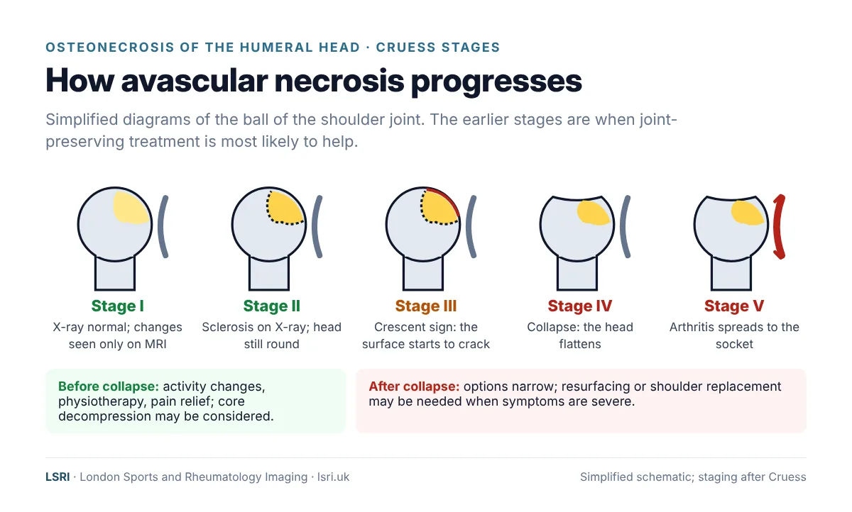
| Stage | What it means | Seen on |
|---|---|---|
| I | Bone has died but the shape of the head is normal | MRI only; X-ray normal |
| II | Sclerosis (denser bone) or thinning, with no collapse | X-ray and MRI |
| III | The surface starts to crack under the joint (the “crescent sign”) | X-ray and MRI |
| IV | The head collapses and flattens | X-ray and MRI |
| V | Arthritis spreads to the socket (glenoid) | X-ray and MRI |
MRI is the most sensitive test for osteonecrosis, particularly in early cases when the X-ray is still normal.3 Left alone, many cases progress. In a long-term study of 215 shoulders with steroid-related osteonecrosis followed for 10 to 20 years, 74% of shoulders that had no symptoms at diagnosis became painful and 54% collapsed; of shoulders that were already painful at diagnosis, 82% went on to collapse.10
Why early diagnosis changes the options
Treatment depends heavily on the stage:4
- Before collapse (stages I–II): pain relief, activity changes and physiotherapy. Core decompression, a procedure that relieves pressure inside the bone, may be considered; a review reported good results in at least 88% of shoulders at stages I and II, falling to about 70% at stage III and as low as 14% at stage IV.
- After collapse (stages III–V): when symptoms are severe, surgery such as resurfacing, partial shoulder replacement (hemiarthroplasty) or total shoulder replacement may be needed.
In other words, a diagnosis made while the ball of the joint is still round keeps joint-preserving options open. A diagnosis made after collapse often does not. That is the real cost of a delayed diagnosis, and it is far greater than the cost of a scan.
Shoulder osteoarthritis: the other half of the picture
Glenohumeral osteoarthritis is wear of the cartilage in the main shoulder joint. It usually affects people over 60, causes pain that is often severe, and leads to stiffness that builds over years, especially loss of the ability to rotate the arm outwards with the elbow by the side.11 UK guidance says it is diagnosed with a plain X-ray, ideally with two views, and lists avascular necrosis of the humeral head among the conditions to consider.11 When osteonecrosis leads to collapse, the uneven joint surface can itself cause arthritis, which is stage V above.
Because osteoarthritis and AVN are problems of bone and cartilage, they will not be resolved by treatments aimed only at the soft tissues. Knowing the diagnosis allows treatment to be planned properly, whether that is targeted physiotherapy, pain management, image-guided injection, or a timely surgical opinion.
When should persistent shoulder pain be scanned?
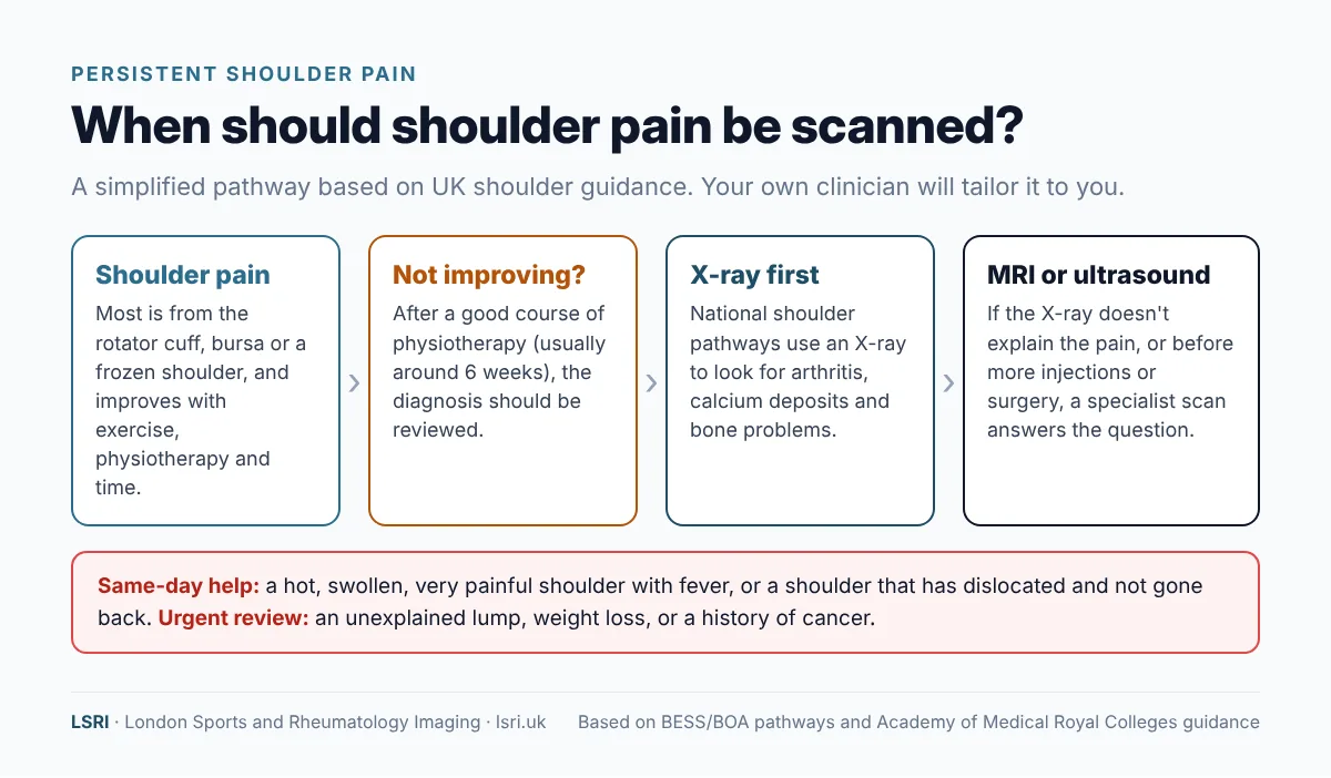
UK guidance offers a clear framework:
- The British Elbow and Shoulder Society and British Orthopaedic Association pathway for subacromial shoulder pain says that two-view shoulder X-rays can be useful when a patient is not improving with conservative treatment, and that conservative care, usually including about six weeks of physiotherapy and no more than two steroid injections, should be followed by referral if it fails.8
- The Academy of Medical Royal Colleges’ Evidence-Based Interventions programme recommends X-ray as the routine first imaging test for most shoulder problems, including arthritis, calcium deposits and tumours, with ultrasound and MRI used in specialist settings.12
- The glenohumeral osteoarthritis pathway recommends referral when pain is not controlled, when stiffness limits function, or when there is any doubt about the diagnosis.11
Put simply: if your shoulder pain has not improved after a good course of physiotherapy, or keeps coming back after injections, ask whether an X-ray has been done and whether the diagnosis should be reviewed. If the X-ray does not explain the pain, or before further injections or surgery, an MRI or ultrasound reported by a musculoskeletal specialist can answer the question. Our guide to MRI or ultrasound for joint pain explains which scan suits which problem.
When shoulder pain needs urgent help
- Same day: a hot, swollen, very painful shoulder, especially with fever or feeling unwell (possible infection), or a shoulder that has dislocated and not gone back.
- Urgently: an unexplained lump or swelling, unexplained weight loss, or shoulder pain in someone with a history of cancer.
Contact NHS 111, your GP or A&E as appropriate.8
How LSRI can help
- The right test first. We offer X-ray, ultrasound and MRI, so we can recommend the scan that answers your question rather than the one that happens to be available.
- Specialist reporting. Your MRI is reported by a consultant musculoskeletal radiologist within 24 hours.
- Diagnosis before more injections. If you have had several injections without lasting benefit, we can review the diagnosis before any further treatment. If an injection is appropriate, it can be guided precisely by ultrasound; see our shoulder injections page.
- Clear pricing. MRI from £375 per body part, including the scan, a specialist report and access to your images, or from £425 with a consultation to go through the results.
Related conditions: frozen shoulder, shoulder impingement, rotator cuff tear and calcific tendinitis. For avascular necrosis of the hip, see our guide to how hip osteonecrosis is diagnosed.
Frequently asked questions
Can avascular necrosis of the shoulder heal?
Dead bone is not easily restored, but early-stage osteonecrosis does not always progress, and symptoms can often be managed. Treatment aims to relieve pain, protect the joint and, where possible, prevent collapse. The earlier it is found, the more options there are.
Can a steroid injection cause avascular necrosis?
The clear link is with high-dose or long courses of steroid tablets or drips. For injections into or around a joint, the evidence is limited to case reports and small studies, and a causal link has not been proven. A scan cannot show the cause in an individual patient, and your specialist will consider all possible risk factors.
Will an X-ray show avascular necrosis?
Not in its earliest stage. In stage I the X-ray is normal and changes are visible only on MRI. Later stages do show on X-ray.
What is the difference between frozen shoulder and shoulder arthritis?
Both cause pain and stiffness. A frozen shoulder is inflammation and tightening of the joint capsule, usually in people aged around 40 to 60, and it typically improves over time. Osteoarthritis is wear of the joint cartilage, usually after 60, and is seen on X-ray. Avascular necrosis can mimic both. See our frozen shoulder page.
How many steroid injections are safe in the shoulder?
There is no single safe number, but UK guidance suggests no more than two as part of conservative care for subacromial pain before referral, and Versus Arthritis advises usually no more than three into the same area in a year. If injections are not giving lasting relief, it is time to confirm the diagnosis.
Book an appointment
Call 0203 633 5040 or book online. Clinics at Harley Street (W1G 9QJ) and Harrow (HA2 7SJ).
References
- Hernigou P, Hernigou J, Scarlat M. Shoulder osteonecrosis: pathogenesis, causes, clinical evaluation, imaging, and classification. Orthopaedic Surgery 2020;12(5):1340–1349. doi:10.1111/os.12788
- Kaza E, Neel G, Feeley S, et al. Humeral head avascular necrosis: etiology, diagnosis, and management. Clinics in Shoulder and Elbow 2025;28(4):517–528. doi:10.5397/cise.2025.00493
- Castillo Mercado JS, Rojas Lievano J, Zaldivar B, et al. Atraumatic osteonecrosis of the humeral head: pathophysiology and current concepts of evaluation and treatment. JSES Reviews, Reports, and Techniques 2022;2(3):277–284. doi:10.1016/j.xrrt.2022.02.005
- Scheidt MD, Aiyash S, Salazar D, et al. Core decompression for early-stage avascular necrosis of the humeral head: current concepts and techniques. Clinics in Shoulder and Elbow 2023;26(2):191–204. doi:10.5397/cise.2022.00969
- Sun W, Shi Z, Gao F, et al. The pathogenesis of multifocal osteonecrosis. Scientific Reports 2016;6:29576. doi:10.1038/srep29576
- Motta F, Timilsina S, Gershwin ME, et al. Steroid-induced osteonecrosis. Journal of Translational Autoimmunity 2022;5:100168. doi:10.1016/j.jtauto.2022.100168
- Zhao G, Tieu PTM, Lameire DL, et al. Avascular necrosis or rapid destruction of the hip following hip intra-articular corticosteroid injections: a systematic review. International Orthopaedics 2025;49(1):127–134. doi:10.1007/s00264-024-06377-0
- Kulkarni R, Gibson J, Brownson P, et al. Subacromial shoulder pain. Shoulder & Elbow 2015;7(2):135–143. doi:10.1177/1758573215576456
- Versus Arthritis. Steroid injections. versusarthritis.org
- Hernigou P, Flouzat-Lachaniette CH, Roussignol X, et al. The natural progression of shoulder osteonecrosis related to corticosteroid treatment. Clinical Orthopaedics and Related Research 2010;468(7):1809–1816. doi:10.1007/s11999-009-1094-1
- Thomas M, Bidwai A, Rangan A, et al. Glenohumeral osteoarthritis. Shoulder & Elbow 2016;8(3):203–214. doi:10.1177/1758573216644183
- Academy of Medical Royal Colleges. Evidence-Based Interventions: scans for shoulder pain and guided injections for shoulder pain. ebi.aomrc.org.uk
About this case study
Patient privacy: the images are anonymised, with no names, dates or other identifying details, and non-essential details of the case have been left out. They are shared for education. Purpose: written by the LSRI clinical team to help the public understand when persistent shoulder pain needs a closer look, based on the research and UK guidance listed above. It is general information, not a diagnosis; speak to your GP or specialist about your own symptoms. The case is not a criticism of any clinician: physiotherapy and, where appropriate, injections are the usual first treatments for shoulder pain.