Written by: LSRI Editorial Team
Medically reviewed by Dr Prashant Sankaye, Consultant MSK Radiologist ·
Last Updated: October 23, 2026
Read Time: 20 Minutes
Written by the LSRI clinical team and medically reviewed by Dr Prashant Sankaye, Consultant MSK Radiologist, on 22 October 2026.
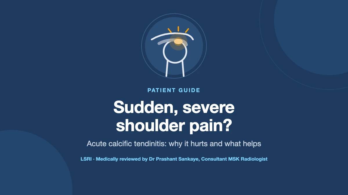
It often starts without warning. A shoulder that was only a little sore becomes so painful over a day or two that you cannot lift your arm, cannot find a comfortable position and cannot sleep. One common cause of this sudden, severe shoulder pain is acute calcific tendinitis: a deposit of calcium in one of the rotator cuff tendons that the body has started to break down. It is also called calcific tendinopathy or calcific tendonitis.
The pain can be frightening, but the news is often better than it feels. This guide, written by the LSRI clinical team led by a consultant musculoskeletal (MSK) radiologist, explains why calcific tendinitis hurts so much, how it is diagnosed, how long it lasts, how to sleep with it, and what the evidence says about injections, barbotage and shockwave therapy.
Key points
- Calcium deposits in the rotator cuff are common. In one study of 1,219 adults, they were found in 7.8% of people with no shoulder pain and 42.5% of people with a common type of shoulder pain (subacromial pain).1
- The severe pain usually comes when the body starts to break the deposit down (the resorptive phase), which raises pressure in the tendon and can inflame the bursa above it.2,3
- An acute flare often settles within one to six weeks, although the calcium itself can take up to nine months, sometimes longer, to clear.4,5
- X-ray and ultrasound are the main tests. MRI is not routine and can make calcium hard to see.2,6
- In a randomised trial, ultrasound-guided barbotage (followed by a steroid injection) gave better results than a steroid injection alone at one year. By five years the results were similar, but fewer people in the barbotage group had needed further treatment.7,8
- The evidence for shockwave therapy comes from long-standing (chronic) calcific tendinitis; it is not well suited to an acute flare.2,9
What is calcific tendinitis?
The rotator cuff is a group of four tendons that hold the ball of the shoulder joint in its socket and help you lift and turn your arm. In calcific tendinitis, a deposit of calcium crystals (mainly calcium hydroxyapatite) forms inside one of these tendons.3 The supraspinatus tendon, at the top of the shoulder, is involved most often.3,10
It is commoner than many people think. Published figures range from about 3% to 20% of adults, depending on who is studied.10 It is most common between the ages of 30 and 60 and about twice as common in women as in men,1,3 and it affects both shoulders in roughly 1 in 10 to 1 in 4 people.3 Many deposits never cause any symptoms at all.1
Why does calcific tendinitis suddenly become so painful?
Calcific tendinitis tends to follow a natural cycle, first described by the Canadian surgeon Hans Uhthoff. He described three stages: a pre-calcific stage, when the tendon tissue changes; a calcific stage, which has the three phases below; and a post-calcific stage, when the tendon repairs.10,11 In simple terms, a deposit forms, rests for a while, and then the body starts to break it down and clear it away:
- Formative phase: the calcium deposit forms in the tendon. It may cause no pain, or an ache with overhead movements.
- Resting phase: the deposit stays as it is, sometimes for years.
- Resorptive phase: the body starts to dissolve the deposit. This is when most people get severe pain.3,10 The deposit softens, the tendon around it swells and the pressure inside the tendon rises, and calcium crystals can leak into the bursa (a fluid-filled cushion) above it, causing intense inflammation.2 The pain can be “highly disabling”.2
- Post-calcific stage: the calcium has gone and the tendon remodels and heals.10
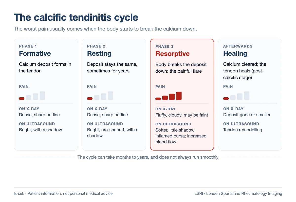
This is why the worst pain is often a sign that the body has started to clear the calcium. Doctors describe the overall process as a self-healing cycle,3 although it does not always run smoothly, and some deposits stay or grow.6
What causes calcific tendinitis?
The exact cause is not known.5 The main theory is that it is an active process driven by tendon cells, a kind of failed healing response, rather than simple wear and tear.3 A few things are linked with it:
- Diabetes: in one controlled study, shoulder calcification was found in 31.8% of adults with diabetes, compared with 10.3% of people without it.12
- Thyroid and hormone-related conditions: in an Oxford study, people with thyroid or oestrogen-related disorders developed symptoms younger and had them for longer.13
- Being a woman aged 30 to 60.1
These are links, not proven causes. There is no evidence that calcium in your diet causes the deposits. In our clinical view, there is no need to cut down on milk, dairy foods or calcium supplements because of calcific tendinitis.
Is it calcific tendinitis, or something else?
Several conditions can cause sudden, severe shoulder pain, and some need urgent treatment. A joint infection (septic arthritis) can look very like an acute calcific attack,6 and pain in the shoulder or arm can come from the heart.14
When shoulder pain needs urgent help
Call 999 or go to A&E if you have shoulder or arm pain with chest pain, shortness of breath, palpitations or pain spreading into your jaw, or if your shoulder looks deformed after a fall or injury.14
Go to A&E straight away if your shoulder is suddenly swollen, red and warm and you have a fever, as this can be a sign of a joint infection.14
Ask for an urgent GP appointment or contact NHS 111 if you cannot move your arm, or you feel feverish or unwell with shoulder pain.15
See your GP urgently if both shoulders (and perhaps your hips) are painful with a lot of morning stiffness.14
Other causes of shoulder pain include a rotator cuff tear, frozen shoulder, arthritis and pain referred from the neck. A scan, read alongside your symptoms and an examination, is the quickest way to tell them apart.
How is calcific tendinitis diagnosed?
The diagnosis is made by putting your symptoms together with imaging. As MSK radiologists, these are the tests we find most useful:
X-ray
Calcium shows up well on X-ray. A deposit that is forming or resting usually looks dense with a sharp outline. In the resorptive phase it often looks fluffy or cloudy, and it can be barely visible.2,6 Radiologists often describe these appearances using the Gärtner classification, from type I (dense and well defined) to type III (cloudy and faint, typical of resorption).2
Ultrasound
Ultrasound shows exactly where the calcium is in the tendon, whether the bursa is inflamed, and whether there is increased blood flow around the deposit, which is seen in the active, resorbing phase.10 “Hard” deposits are bright and cast a dark shadow; “soft” deposits, which are often resorbing, cast little or no shadow.2 Ultrasound also lets us examine the shoulder while you move it, and it is used to guide treatment such as barbotage or an injection. X-ray and ultrasound complement each other.
When does MRI help?
MRI is not a routine test for calcific tendinitis. Calcium is dark on MRI and easy to miss, and the swelling around it can look like a tendon tear.2,6 MRI is useful when another problem is suspected, such as a rotator cuff tear, or when calcium has spread into the bone, which can look worrying on imaging and needs specialist interpretation.2,16 For more on choosing a scan, see MRI or ultrasound for joint pain?
How long does calcific tendinitis last?
An acute flare often settles by itself within one to six weeks.4 The pain usually eases long before the calcium has gone: the deposit can take up to nine months, or sometimes longer, to dissolve.5 Most cases settle without a procedure,10 and acute cases generally respond well to simple treatment. Long-standing (chronic) cases are more likely to need more help.6
In the longer term, the outlook is generally good. When 35 people were re-examined around 13 years after diagnosis, most deposits had shrunk, although most people still had some calcium visible, and none had a full-thickness rotator cuff tear.17 Deposits do not always shrink, though: in one series, 18% grew over an average of 16 months.6
How to sleep with calcific tendonitis
Night pain is one of the hardest parts of an acute attack, and lying on the painful side usually makes it worse.5 No research trial has tested sleeping positions, so these tips from NHS physiotherapy services may or may not help you. Use whatever is most comfortable:
- Sleep propped up. You may find it more comfortable to sleep propped up on pillows.4
- Support the arm. NHS physiotherapists suggest using a pillow to support the painful arm.14
- Stop yourself rolling over. A pillow can stop you rolling onto the painful side.14
- Time your pain relief. Ask a pharmacist or GP whether you can take pain relief, such as paracetamol or ibuprofen, before bed.15
- Try ice. A pack of frozen peas wrapped in a tea towel for up to 20 minutes can help.15
If you cannot sleep at all despite this, or the pain is getting worse, get it assessed. For some people, treatment such as an ultrasound-guided steroid injection can help control pain in the acute stage, including at night.5
Treatment for calcific tendinitis: what the evidence shows
First steps: pain relief, ice and keeping moving
The NHS advises keeping the shoulder gently moving rather than stopping completely, using ice or heat, and trying pain relief such as paracetamol or ibuprofen.15 Anti-inflammatory medicines are commonly used during an acute flare,10 although there is little trial evidence for them in calcific tendinitis specifically. Shoulder exercises can help in the later stages, once the acute pain has settled,5 but a 2020 review found no randomised trials of exercise for calcific tendinitis.18
Steroid injection
A steroid (corticosteroid) injection into the bursa above the tendon can reduce inflammation and pain in the acute stage.5 Under ultrasound guidance, the needle can be placed precisely. On its own, a steroid injection eases symptoms but does not directly remove the calcium. Some experts are concerned it may slow the body’s resorption of the deposit,10 but in a randomised trial a steroid injection did not significantly change how often the calcium cleared.19
Barbotage (ultrasound-guided lavage)
Barbotage, also called ultrasound-guided needling and lavage, uses a fine needle under ultrasound to break up the deposit and wash out as much calcium as possible with saline. It is usually followed by a steroid injection into the bursa.7
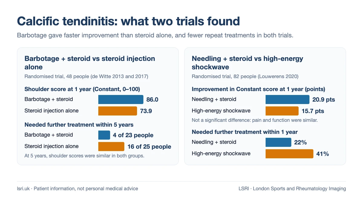
- Compared with a steroid injection alone: in a randomised trial of 48 people (23 had barbotage plus a steroid injection, 25 had the steroid injection alone), the barbotage group had better shoulder scores at one year (86.0 vs 73.9 on the 100-point Constant score) and greater shrinkage of the deposit.7
- At five years: the two groups had similar results, but only 4 of the 23 people in the barbotage group had needed further treatment, compared with 16 of the 25 in the steroid-only group.8 So barbotage seems to speed up recovery and reduce the need for more procedures, rather than give a better final result.
- Why a steroid is added: in another trial, a steroid injection after lavage reduced pain over the first six weeks, improved function for three months, and did not significantly change how often the calcium cleared by 12 months.19
- What to expect afterwards: complications are uncommon and usually minor, such as feeling faint during the procedure or a flare of pain afterwards as the bursa reacts.2,20 Not every deposit can be fully washed out,21 and some people need a repeat procedure.8
Barbotage at LSRI: one patient’s images
These anonymised images come from one of our patients with calcific tendinitis of the shoulder. The MRI before treatment showed a calcium deposit about 2 cm long in the rotator cuff. Calcium looks dark on MRI, which is one reason it can be hard to spot on MRI alone.2,6
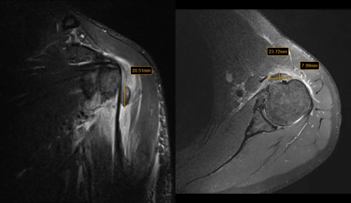
Dr Sankaye then treated the deposit with ultrasound-guided barbotage under local anaesthetic. The short ultrasound recording below is from the procedure.
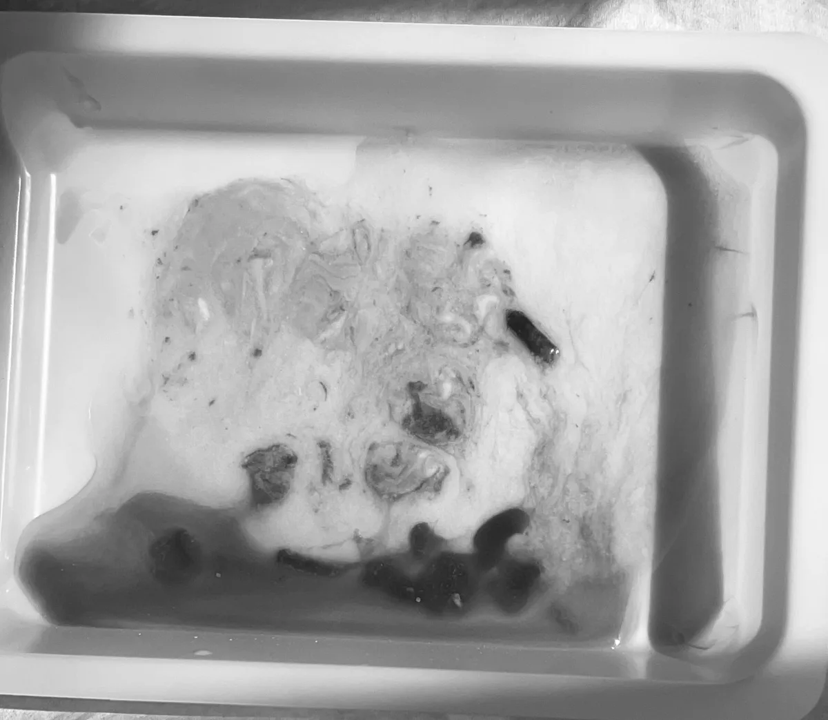
The photo shows the calcium that was washed out of the tendon, collected in a tray. Results vary from person to person: not every deposit can be washed out this well, and some people need a repeat procedure.8,21
Shockwave therapy for calcific tendonitis
Extracorporeal shockwave therapy (ESWT) sends pulses of sound energy through the skin to the deposit. In a sham-controlled trial of long-standing calcific tendinitis, high-energy shockwave aimed at the deposit using X-ray guidance, given in two sessions about two weeks apart, improved Constant scores by 31.0 points at six months, compared with 15.0 for low-energy treatment and 6.6 for sham treatment.9 A systematic review concluded that high-energy shockwave is effective for chronic calcific tendinitis and can clear the deposits.22
The wider picture is more mixed. A Cochrane review of shockwave for rotator cuff problems, including calcific tendinitis, found that the benefits were small and mostly not clinically important, with no clear difference in effect between people with and without calcium deposits.23 Two other points matter if you are considering it. First, most of the good evidence is for high-energy focused shockwave; many clinics use radial devices, which have much less evidence for calcium deposits.24 Second, shockwave is not well suited to the acute, resorptive flare,2 and high-energy treatment can itself be painful.10
Barbotage or shockwave?
In a randomised trial of 82 people comparing ultrasound-guided needling (with a steroid injection) and high-energy shockwave, pain and function were similar at one year. Needling removed more calcium, and fewer people needed further treatment: 22% after needling compared with 41% after shockwave.25
Surgery
Keyhole (arthroscopic) surgery to remove the calcium is usually considered only when symptoms have not improved after at least six months of non-surgical treatment.6,10 Recovery is slow, often six months or more.6
Calcific tendinitis in other places
Calcium deposits can form in other tendons too. Around the hip, the gluteus medius tendon is the most common site, and most cases improve without surgery.26 Rarely, a deposit in a tendon in front of the spine, behind the throat, causes sudden neck pain and pain on swallowing.27
How LSRI can help
LSRI is a doctor-led musculoskeletal (MSK) imaging and injection clinic, not an emergency service. If anything in the red box applies to you, get urgent care first.
- Diagnosis with ultrasound by Dr Prashant Sankaye, Consultant MSK Radiologist, with an X-ray when needed, to find the calcium, see which phase it is in, and rule out other causes. Ultrasound starts from £200 at our Harley Street and Harrow clinics, and X-ray from £200 at our partner imaging centre in Fitzrovia, central London.
- Ultrasound-guided treatment, including shoulder injections and barbotage, when it is the right option for you. See our calcific tendinitis treatment page and prices.
- MRI at our partner imaging centres when another problem needs to be ruled out, in two options: scan only, from £375 per body part (the scan, a report from a consultant MSK radiologist within 24 hours and access to your images, without a consultation or advice on treatment), or scan with a consultation, from £425, which adds a consultation to go through your results and next steps. Our clinical team checks every MRI request first and will tell you if an X-ray or ultrasound would answer your question better; see do you need a GP referral for a private MRI?.
- A second opinion on a shoulder scan you have already had: see our second opinion page.
What patients say about LSRI
“Highly recommend [Dr Sankaye] to assist you with your MSK related matters – they have a fantastic team at LSRI to handle all your questions and concerns.”
A., Google review of LSRI Harrow
“This doctor is an angel, he immediately found where my issue rooted from … Thank you so much and I wish the best for this doctor.”
Patient, Google review of LSRI Harrow
Reviews describe individual experiences of our service; results of treatment vary from person to person.
Our clinics are on Harley Street (W1G 9QJ) and in Harrow (HA2 7SJ); call 0203 633 5040. Read more: shoulder pain not getting better with physio?
Reading this from outside the UK?
The medical points here apply everywhere, but emergency numbers differ: 999 or 112 in the UK, 112 across the EU, 911 in the US and Canada, and 000 in Australia. NHS 111 is a UK service; elsewhere, use your local urgent care service.
Frequently asked questions
Does calcific tendinitis go away on its own?
Usually, yes. Most cases settle without a procedure as the body breaks down the deposit,10 although this can take months, and some deposits stay or grow.6
Why is calcific tendinitis so painful at night?
In the resorptive phase, inflammation and raised pressure in and around the tendon make the shoulder very sensitive,2 and lying on it squeezes the inflamed area. Sleeping propped up with the arm supported on a pillow may be more comfortable; this is NHS advice rather than something tested in trials.4,14
Is barbotage painful?
The area is numbed with local anaesthetic first, so the procedure itself is usually more uncomfortable than painful. A flare of pain afterwards is one of the more common side effects, and usually settles.2 A steroid injection given at the same time reduces pain over the first few weeks.19
Can calcific tendinitis cause a rotator cuff tear?
Long-term follow-up suggests this is uncommon. In a study that re-examined people about 13 years after diagnosis, none had a full-thickness rotator cuff tear.17 Long-standing pain can, however, lead to a stiff shoulder.6
Should I stop taking calcium or eating dairy?
No. There is no evidence that calcium in your diet causes calcific tendinitis. If you take calcium or vitamin D for your bones, keep taking it unless your doctor advises otherwise.
Can calcific tendinitis come back?
Both shoulders are affected in about 1 in 10 to 1 in 4 people,3 and some people need a further procedure after treatment.8,25
References
- Louwerens JK, Sierevelt IN, van Hove RP, van den Bekerom MP, van Noort A. Prevalence of calcific deposits within the rotator cuff tendons in adults with and without subacromial pain syndrome: clinical and radiologic analysis of 1219 patients. J Shoulder Elbow Surg 2015;24(10):1588-93. doi:10.1016/j.jse.2015.02.024
- Chianca V, Albano D, Messina C, Midiri F, Mauri G, Aliprandi A, et al. Rotator cuff calcific tendinopathy: from diagnosis to treatment. Acta Biomed 2018;89(1-S):186-96. doi:10.23750/abm.v89i1-S.7022
- Sansone V, Maiorano E, Galluzzo A, Pascale V. Calcific tendinopathy of the shoulder: clinical perspectives into the mechanisms, pathogenesis, and treatment. Orthop Res Rev 2018;10:63-72. doi:10.2147/ORR.S138225
- York and Scarborough Teaching Hospitals NHS Foundation Trust, Virtual Fracture Clinic. Calcific tendonitis: advice and exercises (patient letter). yorkhospitals.nhs.uk (accessed 22 October 2026)
- NHS Lothian. Calcific tendinopathy: information for patients. Version 2.0, February 2025. nhslothian.scot (accessed 22 October 2026)
- Kim MS, Kim IW, Lee S, Shin SJ. Diagnosis and treatment of calcific tendinitis of the shoulder. Clin Shoulder Elb 2020;23(4):210-6. doi:10.5397/cise.2020.00318
- de Witte PB, Selten JW, Navas A, Nagels J, Visser CP, Nelissen RG, et al. Calcific tendinitis of the rotator cuff: a randomized controlled trial of ultrasound-guided needling and lavage versus subacromial corticosteroids. Am J Sports Med 2013;41(7):1665-73. doi:10.1177/0363546513487066
- de Witte PB, Kolk A, Overes F, Nelissen RGHH, Reijnierse M. Rotator cuff calcific tendinitis: ultrasound-guided needling and lavage versus subacromial corticosteroids: five-year outcomes of a randomized controlled trial. Am J Sports Med 2017;45(14):3305-14. doi:10.1177/0363546517721686
- Gerdesmeyer L, Wagenpfeil S, Haake M, Maier M, Loew M, Wörtler K, et al. Extracorporeal shock wave therapy for the treatment of chronic calcifying tendonitis of the rotator cuff: a randomized controlled trial. JAMA 2003;290(19):2573-80. doi:10.1001/jama.290.19.2573
- Merolla G, Singh S, Paladini P, Porcellini G. Calcific tendinitis of the rotator cuff: state of the art in diagnosis and treatment. J Orthop Traumatol 2016;17(1):7-14. doi:10.1007/s10195-015-0367-6
- Uhthoff HK, Loehr JW. Calcific tendinopathy of the rotator cuff: pathogenesis, diagnosis, and management. J Am Acad Orthop Surg 1997;5(4):183-91. doi:10.5435/00124635-199707000-00001
- Mavrikakis ME, Drimis S, Kontoyannis DA, Rasidakis A, Moulopoulou ES, Kontoyannis S. Calcific shoulder periarthritis (tendinitis) in adult onset diabetes mellitus: a controlled study. Ann Rheum Dis 1989;48(3):211-4. doi:10.1136/ard.48.3.211
- Harvie P, Pollard TC, Carr AJ. Calcific tendinitis: natural history and association with endocrine disorders. J Shoulder Elbow Surg 2007;16(2):169-73. doi:10.1016/j.jse.2006.06.007
- Leicestershire Partnership NHS Trust, MSK Physiotherapy Service. Shoulder pain: patient information leaflet. April 2026. leicspart.nhs.uk (accessed 22 October 2026)
- NHS. Shoulder pain. nhs.uk (page last reviewed 22 May 2023; accessed 22 October 2026)
- Flemming DJ, Murphey MD, Shekitka KM, Temple HT, Jelinek JJ, Kransdorf MJ. Osseous involvement in calcific tendinitis: a retrospective review of 50 cases. AJR Am J Roentgenol 2003;181(4):965-72. doi:10.2214/ajr.181.4.1810965
- Compagnoni R, Menon A, Radaelli S, Lanzani F, Gallazzi MB, Tassi A, et al. Long-term evolution of calcific tendinitis of the rotator cuff: clinical and radiological evaluation 10 years after diagnosis. J Orthop Traumatol 2021;22(1):42. doi:10.1186/s10195-021-00604-9
- Simpson M, Pizzari T, Cook T, Wildman S, Lewis J. Effectiveness of non-surgical interventions for rotator cuff calcific tendinopathy: a systematic review. J Rehabil Med 2020;52(10). doi:10.2340/16501977-2725
- Darrieutort-Laffite C, Varin S, Coiffier G, Albert JD, Planche L, Maugars Y, et al. Are corticosteroid injections needed after needling and lavage of calcific tendinitis? Randomised, double-blind, non-inferiority trial. Ann Rheum Dis 2019;78(6):837-43. doi:10.1136/annrheumdis-2018-214971
- Lanza E, Banfi G, Serafini G, Lacelli F, Orlandi D, Bandirali M, et al. Ultrasound-guided percutaneous irrigation in rotator cuff calcific tendinopathy: what is the evidence? A systematic review with proposals for future reporting. Eur Radiol 2015;25(7):2176-83. doi:10.1007/s00330-014-3567-1
- Oudelaar BW, Huis In ‘t Veld R, Schepers-Bok R, Ooms EM, Nelissen RGHH, Vochteloo AJH. Prognostic factors for the outcome of needle aspiration of calcific deposits for rotator cuff calcific tendinitis. Eur Radiol 2020;30(7):4082-90. doi:10.1007/s00330-020-06669-0
- Bannuru RR, Flavin NE, Vaysbrot E, Harvey W, McAlindon T. High-energy extracorporeal shock-wave therapy for treating chronic calcific tendinitis of the shoulder: a systematic review. Ann Intern Med 2014;160(8):542-9. doi:10.7326/M13-1982
- Surace SJ, Deitch J, Johnston RV, Buchbinder R. Shock wave therapy for rotator cuff disease with or without calcification. Cochrane Database Syst Rev 2020;3(3):CD008962. doi:10.1002/14651858.CD008962.pub2
- de Boer FA, Mocking F, Nelissen EM, van Kampen PM, Huijsmans PE. Ultrasound guided needling vs radial shockwave therapy in calcific tendinitis of the shoulder: a prospective randomized trial. J Orthop 2017;14(4):466-9. doi:10.1016/j.jor.2017.07.011
- Louwerens JKG, Sierevelt IN, Kramer ET, Boonstra R, van den Bekerom MPJ, van Royen BJ, et al. Comparing ultrasound-guided needling combined with a subacromial corticosteroid injection versus high-energy extracorporeal shockwave therapy for calcific tendinitis of the rotator cuff: a randomized controlled trial. Arthroscopy 2020;36(7):1823-33.e1. doi:10.1016/j.arthro.2020.02.027
- Park SM, Baek JH, Ko YB, Lee HJ, Park KJ, Ha YC. Management of acute calcific tendinitis around the hip joint. Am J Sports Med 2014;42(11):2659-65. doi:10.1177/0363546514545857
- Horowitz G, Ben-Ari O, Brenner A, Fliss DM, Wasserzug O. Incidence of retropharyngeal calcific tendinitis (longus colli tendinitis) in the general population. Otolaryngol Head Neck Surg 2013;148(6):955-8. doi:10.1177/0194599813482289
Book an appointment
Call 0203 633 5040 or book online. Clinics at Harley Street (W1G 9QJ) and Harrow (HA2 7SJ). If you have emergency symptoms, go to A&E or call 999 instead.
About this article
Purpose: written by the LSRI clinical team to explain acute calcific tendinitis of the shoulder: why it causes sudden, severe pain, how it is diagnosed, how long it lasts, how to manage night pain, and what the evidence shows for injections, barbotage, shockwave therapy and surgery, based on the research and guidance listed above. It is general information, not a diagnosis. If you are worried about your own symptoms, speak to your GP or, in an emergency, go to A&E.