Written by: LSRI Editorial Team
Medically reviewed by Dr Prashant Sankaye, Consultant MSK Radiologist ·
Last Updated: October 14, 2026
Read Time: 20 Minutes
Written by the LSRI clinical team and medically reviewed by Dr Prashant Sankaye, Consultant MSK Radiologist, on 11 October 2026.
Can an MRI miss something? It is a question we are often asked, usually by people whose pain does not match what their scan report says. The honest answer is: yes, sometimes. But it is rarely the MRI machine that “misses”. Most findings that are missed were there on the images. They were not recognised, were not looked for, or the scan was not set up to show them clearly.
Errors in reading scans are a well-studied part of radiology everywhere in the world, and understanding why they happen is the best way to reduce them. This guide explains how often scan findings are missed, the common reasons, and what you can do to lower the risk for your own scan.
Key points
- Most missed findings on scans are reading (interpretation) errors rather than failures of the machine, and the overall rate has changed little in decades.1
- In day-to-day practice, radiologists’ error rate is estimated at around 3 to 5% of scans reported.2,3 Many of these errors are minor, but some matter.2
- The most common reasons are under-reading and “satisfaction of search”: stopping looking once one abnormality has been found.4
- Missing or inaccurate clinical information is a common and avoidable problem. UK reporting standards state that interpretation “may be incorrect if referral details are deficient, absent or misleading”.5
- In one study, a short conversation between the radiologist and the patient led to changes in 42.6% of MRI reports, almost all because clinical information had been missing.6
- You can lower the risk: give a clear history, bring previous scans, ask who will report your scan, and ask for a review if your report does not match your symptoms.
Can an MRI miss something? Yes, but it is usually not the machine
A modern MRI scanner produces hundreds of detailed images of your body. Whether those images lead to the right diagnosis depends on three things:
- How the scan was set up (the “protocol”): which part of the body was scanned, which sequences were used, in which planes and at what slice thickness. A small tear or an early stress fracture can be hard to see if the scan was not designed to look for it.
- How the images were read: whether the radiologist saw the abnormality (perception) and then understood what it meant (interpretation).
- What the radiologist knew about you: where it hurts, how it started, what you have already tried, and what has happened to you before.
Reviews of error in radiology conclude that most missed radiological diagnoses come down to how the images were interpreted, and that the overall rate of error does not appear to have changed for more than 50 years.1 Better scanners alone have not solved the problem, because the problem is mostly human and organisational.
How common are reporting errors?
Radiologists have studied their own mistakes since the 1940s, and two figures come up again and again:2,7
- About 3 to 5% in everyday practice. Across all scans reported in a normal working day, the real-time error rate is estimated at about 3 to 5%. Many of these are minor and do not change treatment.2
- About 30% in hindsight. When scans that are known to be abnormal are looked at again, around 30% of the abnormalities turn out to have been missed the first time.2 This does not mean 30% of reports are wrong. It means that subtle abnormalities are easy to miss, and much easier to see once you know they are there.
Because so many scans are performed, even a small percentage adds up: one review estimated that there are around 40 million diagnostic errors involving imaging worldwide every year.3 Errors are usually grouped into two types. Perceptual errors, where the abnormality is on the images but is not seen, make up an estimated 60 to 80%. Cognitive or interpretive errors, where the abnormality is seen but its meaning is misjudged, make up the remaining 20 to 40%.2
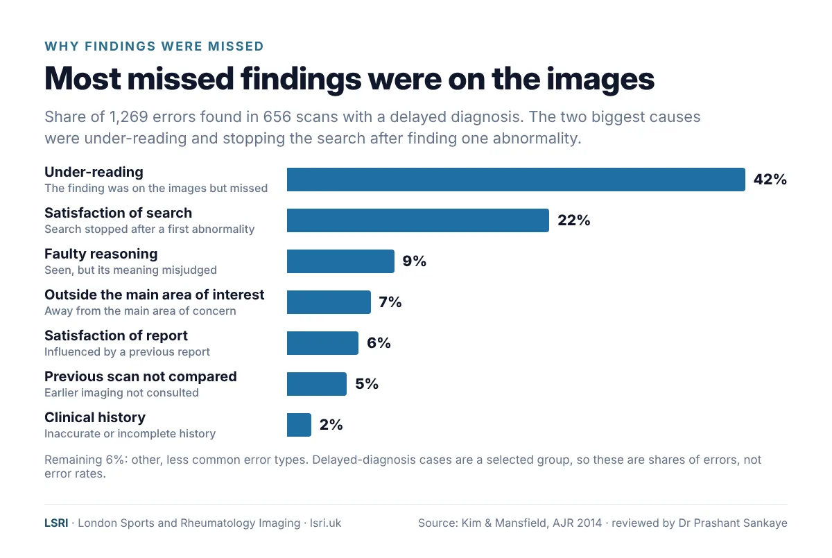
In that study of delayed diagnoses, the correct diagnosis was also missed on a later scan in 30% of cases.4 Once a report says “normal”, the next reader can be influenced by it. That is one reason a good second opinion looks at the images afresh, rather than simply re-reading the earlier report.
Seven common reasons scan findings are missed
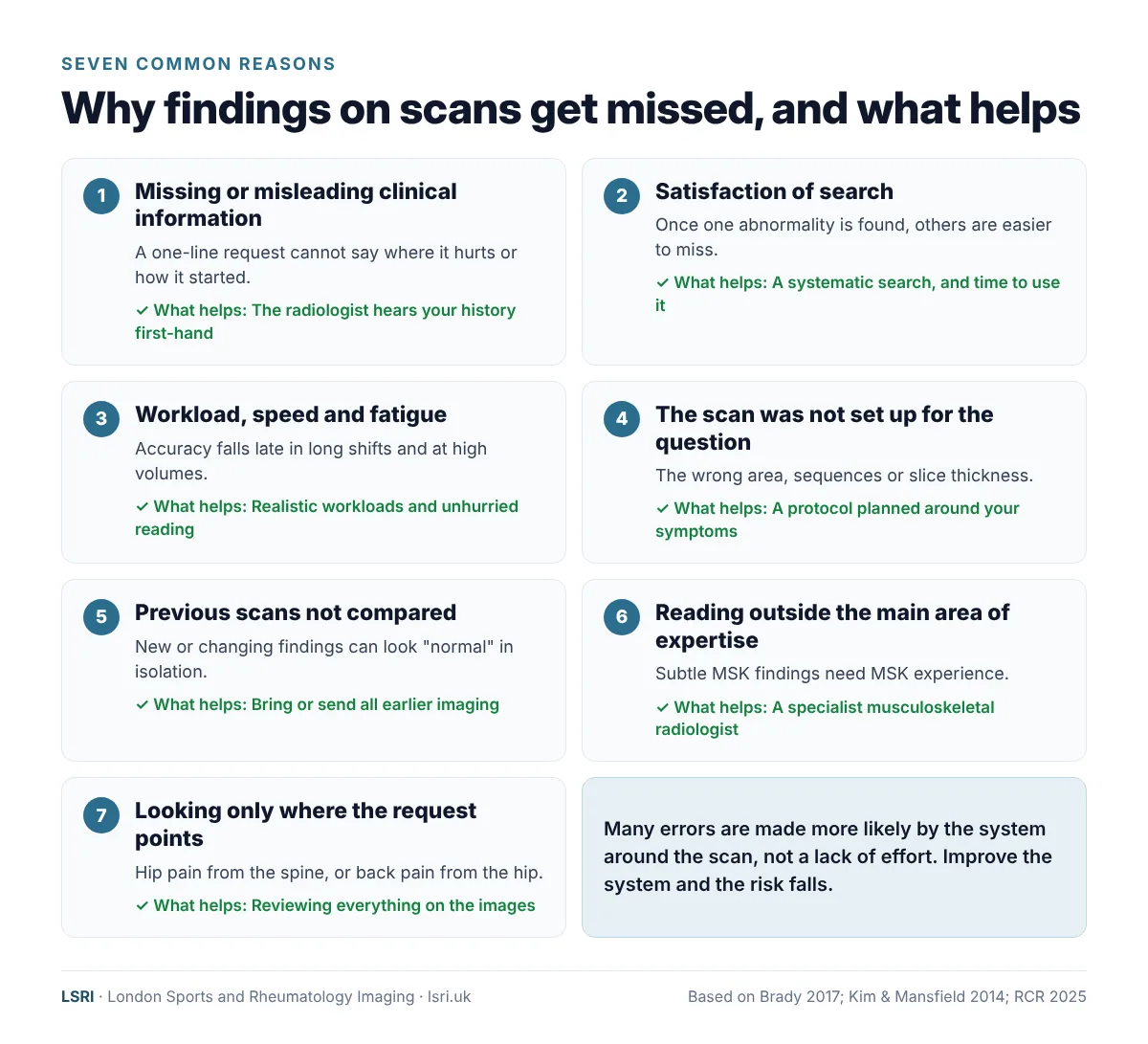
1. Missing or misleading clinical information
A radiologist reads your scan in the light of the question they have been asked. If the request says only “knee pain”, the radiologist does not know whether the pain started with a twisting injury, has come on slowly over a year, wakes you at night, or follows an operation. Each of these points the search in a different direction.
Request forms are often brief. In a US study of lumbar spine MRI and abdominal CT requests, the clinical information was incomplete in 81% when compared with the clinician’s own notes.8 In an evaluation of 1,548 acute imaging requests at a UK hospital, 76% asked a specific clinical question, and where a patient had relevant past medical or surgical history, it was mentioned in only 49%.9
The evidence that good clinical information helps is consistent. A systematic review in JAMA found that, overall, clinical information improved the accuracy of reading tests, and there were no instances in which it significantly reduced accuracy.10 A later review of 20 studies reached a similar conclusion.11 In musculoskeletal imaging specifically, telling radiologists exactly where the pain, tenderness or swelling was improved their detection of subtle fractures, without increasing false alarms.12 In another study, simply marking the site of symptoms on a diagram of the foot increased radiologists’ sensitivity for subtle fractures on foot X-rays from 67% to 73%.13
The UK’s Royal College of Radiologists puts it plainly: “High-quality image interpretation starts with high-quality referrals”, and interpretation “may be incorrect if referral details are deficient, absent or misleading”.5
In the delayed-diagnosis study above, poor clinical history was recorded as the main cause in only 2% of errors, but missing information often contributes alongside other causes, and studies that add clinical information consistently show benefits. This is not a criticism of the clinicians who refer patients: request forms have limited space, and no referrer can know every detail the radiologist will need. Hearing it from you directly fills the gaps.
2. Satisfaction of search
Once the eye finds one abnormality, it is natural to feel the job is done. Radiologists call this satisfaction of search, and it was the second most common cause of error in the study above (22%).4 It happens in bone and joint imaging too: in an experiment using musculoskeletal X-rays, readers found the first abnormality just as well in cases with several findings, but were significantly worse at detecting the second and third.14 The cure is a systematic search pattern, and enough time to use it.
3. Workload, speed and fatigue
Reading scans is intense visual and mental work. In one experiment, radiologists’ accuracy at detecting fractures on bone X-rays fell after a long day of reading, and they reported more fatigue and eye strain.15 In an analysis of 2.9 million examinations from a large US radiology practice, significant reporting errors were more common late in long shifts, peaking 10 to 12 hours in, and on shifts with much higher volumes.16 In a small pilot study, radiologists asked to read abdominal CT scans faster made more major misses than when reading at their normal speed.17
This is a system problem, not a matter of individual effort. In the Royal College of Radiologists’ 2025 census, only around one in five (19%) radiology clinical directors said they had enough consultant radiologists for safe and effective patient care.18
4. The scan was not set up for the question
An MRI scan is not a single photograph. Each examination is built from several sequences, each showing tissues in a different way, and each acquired in particular planes and slice thicknesses. Guidance on knee MRI notes that many practices tailor each study to the specific clinical question, and that it is the supervising doctor’s responsibility to decide whether additional or specialised sequences are needed.19 If the wrong area is scanned, the slices are too thick, or a key sequence is missing, a finding can be invisible however carefully the images are read. UK standards expect the person reporting the scan to be able to judge the quality of the images.5
5. Previous scans were not compared
Comparing with earlier imaging shows whether something is new, growing or long-standing. Not consulting a previous examination accounted for 5% of errors in the study of delayed diagnoses.4 UK standards say a patient’s imaging history and reports should be available to the person reporting the scan.5
6. The scan is read outside the reporter’s main area of expertise
Radiology covers the whole body, and most radiologists specialise further. Studies comparing reports by musculoskeletal subspecialists with reports by radiologists without that subspecialty training have found differences in accuracy and in clinically significant findings, particularly in complex areas such as the hip and bone and soft-tissue tumours.20,21 We explain this in more detail in our article on why it matters who reports your MRI.
7. Looking only where we were asked to look
An abnormality outside the main area of interest accounted for 7% of errors in the delayed-diagnosis study, and being influenced by a previous report accounted for 6%.4 Back pain that is really coming from the hip, or hip pain coming from the spine, is a classic musculoskeletal example. A good report looks at everything on the images, not only the part named on the request.
Why talking to the patient matters so much in musculoskeletal imaging
In bone, joint and spine imaging, the scan often shows several “abnormalities”, and the real question is which one is causing your symptoms. Many changes are a normal part of getting older. A systematic review found disc degeneration on the scans of 37% of 20-year-olds and 96% of 80-year-olds who had no back pain at all. The authors concluded that these findings “must be interpreted in the context of the patient’s clinical condition”.22
That context is best heard from you. In a study of 122 outpatients having MRI, radiologists first reported each scan using only the information on the request; a radiologist then spoke to the patient for about three minutes, and the scan was read again. After the conversation, 52 reports (42.6%) were changed, and in 96.2% of these the reason was clinical information that had been missing from the request.6 In another study, six musculoskeletal radiologists reading lumbar spine MRI agreed far more closely with spine specialists about the likely source of pain when they had the patient’s own symptom questionnaire than when they did not.23
Accuracy of the information matters as much as its presence: in one study, clinical information made CT reports more accurate when it was correct and less accurate when it was wrong.24 Hearing the history directly from the patient, rather than second-hand through a request form, is one way to make it more accurate. This is why at LSRI the radiologist talks to you. We explain how this works in our article on the radiology consultation.
What you can do to reduce the risk of something being missed
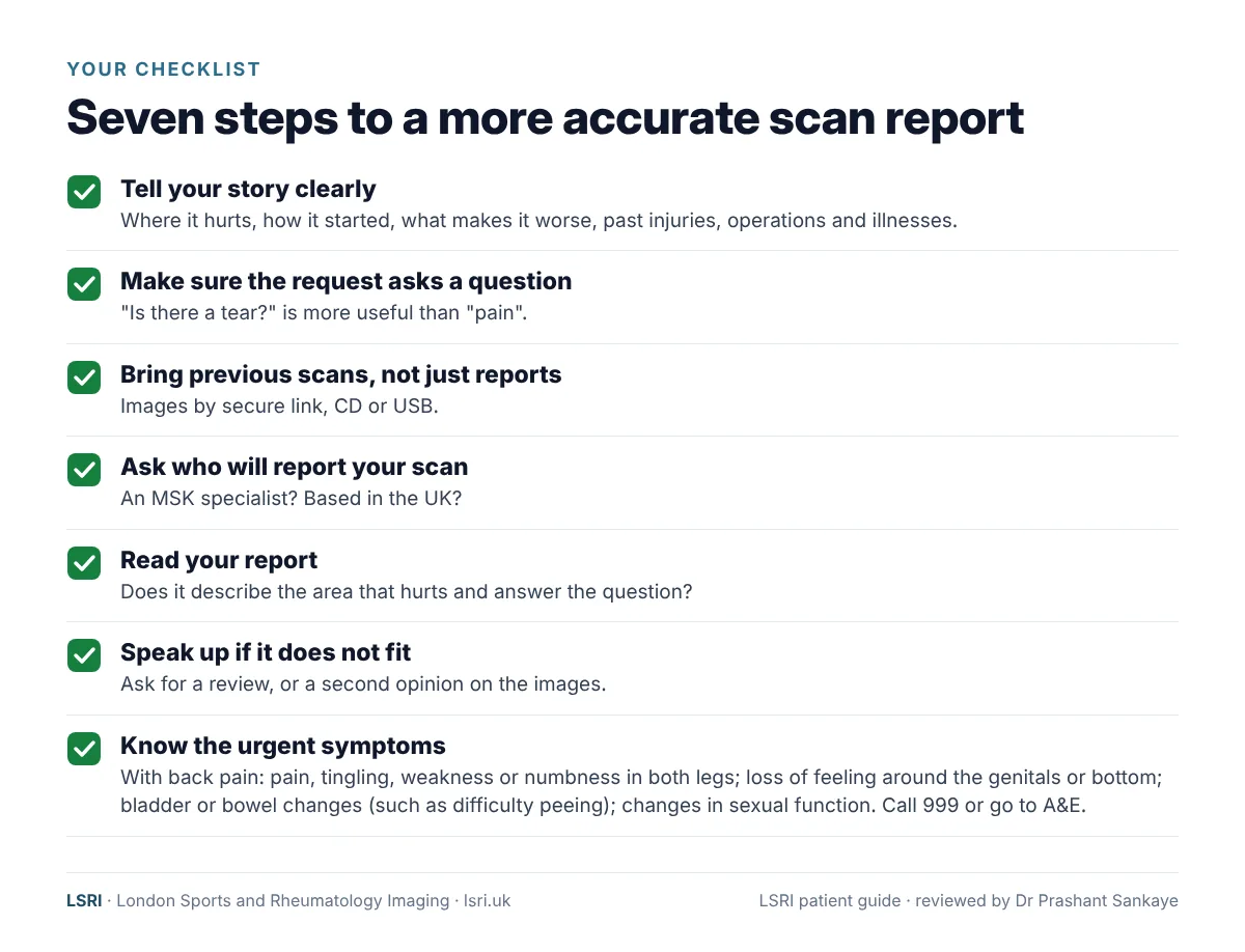
- Tell your story clearly. Where exactly is the pain? When and how did it start? What makes it better or worse? Have you had injuries, operations, cancer, arthritis or other conditions before? Write it down and bring it with you.
- Make sure the request asks a question. “Is there a tear?” or “Could this be a stress fracture?” is far more useful than “pain”.
- Bring previous scans, not just reports. Images can usually be shared by secure link, CD or USB. Comparing with earlier scans is one of the most powerful tools a radiologist has.
- Ask who will report your scan. Is the radiologist a specialist in musculoskeletal imaging, and are they based in the UK? Remote reporting services, especially if based outside the UK, may not have the full context of your case.
- Read your report. Does it describe the area that hurts? Does it answer the question that was asked? If you do not understand it, ask.
- If your symptoms and your report do not match, say so. Ask your clinician to review the diagnosis, and consider a second opinion on the images.
- Know when it is urgent. If you have back pain with pain, tingling, weakness or numbness in both legs, loss of feeling around your genitals or bottom, changes in your bladder or bowels (such as difficulty peeing, or peeing or pooing yourself), changes in sexual function, or chest pain, or if it started after a serious accident, call 999 or go to A&E.25
When is a second opinion worth considering?
Second opinions are an accepted part of good medical practice. A systematic review of patient-initiated second opinions found that between 10% and 62% led to a major change in diagnosis, treatment or prognosis.26 In musculoskeletal imaging, a systematic review of second-opinion reporting by specialist musculoskeletal radiologists found that clinically significant differences ranged from 1.4% to 27.9%, lowest for routine X-rays and knee MRI and highest for tumours and complex cases.27 In other words, many second opinions confirm the original report, and that reassurance is valuable in itself. But in complex cases, a specialist second look changes the picture surprisingly often. A second opinion is worth considering if:
- your symptoms do not fit what your report says, or have not improved as expected;
- you are facing surgery, an injection or a long period off work or sport on the strength of a scan;
- your report is unclear, or different reports disagree;
- a lump, bone lesion or possible tumour has been found;
- the scan is important for an insurance, employment or legal decision.
A second opinion should never delay urgent care. If a possible tumour has been found, keep any appointments your specialist team has arranged and tell them you are seeking a second opinion.
Find out more about our MRI second opinion service.
How we work at LSRI
No radiologist can promise never to miss anything, and we do not. What we can do is build our service around the things that are known to reduce error:
- We listen first. Before your scan, we take your history. Dr Prashant Sankaye, Consultant MSK Radiologist, reads it alongside your images and, if anything is unclear, may contact you before the report is finalised.
- Musculoskeletal specialists only. Every LSRI report is written by a UK-governed consultant musculoskeletal radiologist.
- The scan is set up for your question. The protocol is chosen around your symptoms and your question.
- Previous imaging is compared whenever you can share it.
- A report that answers your question, with a clear summary, and time to talk it through.
“No radiologist can say they will never miss anything. But taking the time to talk to the patient and understand their problem helps me focus on what really matters to them. Radiology is a clinical specialty: the patient comes first, not just the pictures.”
Dr Prashant Sankaye, Consultant MSK Radiologist
How LSRI can help
- Advice before you book. Call us to discuss whether a scan is likely to help, and which one.
- Specialist reporting. Your MRI is reported by a consultant musculoskeletal radiologist within 24 hours.
- Clear pricing. MRI from £375 per body part, including the scan, a specialist report and access to your images.
- Time to talk. MRI with a consultation, to go through the results and next steps, is from £425.
- Second opinions. Already had a scan elsewhere? We can review the images and give you an independent second opinion.
You may also find these useful: Is a cheap MRI scan worth it? and understanding your MRI scan results.
Frequently asked questions
Can an MRI miss a tear?
Yes, especially small or partial tears. Partial-thickness rotator cuff tears in the shoulder are a well-known example: in an older (1997) study, radiologists of varying experience detected only 35 to 44% of them on MRI.28 Scanners and techniques have improved since, but small partial tears remain among the harder diagnoses. The right sequences, a clear clinical question and a specialist reader all help, and sometimes ultrasound, which can be done while you move the joint, adds information.
Can an MRI miss a fracture?
MRI is the most sensitive routine test for many fractures, including stress fractures that do not show on X-ray. But very subtle fractures can still be missed, particularly if the area was not fully covered or the right sequences were not used. See our guide to stress fractures that don’t show on X-ray.
Is it normal for two radiologists to report the same scan differently?
Some variation is normal, particularly in how findings are described and graded. In one illustrative study, the same patient had lumbar spine MRI at 10 different centres within three weeks: 49 different findings were reported, and not one was reported by all 10 centres.29 That was a single patient, so it cannot tell us how common such variation is, but it shows why who reads your scan, and what they know about you, matters.
Can I ask for my scan to be reviewed?
Yes. You can ask your referring clinician to discuss it with the radiologist, or you can ask for an independent second opinion. You can usually request a copy of your images and report from the hospital or clinic where you were scanned.
Will my doctor mind if I get a second opinion?
Second opinions are a normal part of medicine, and most clinicians welcome more information. With your permission, we can send our report to your GP, consultant or physiotherapist.
Book an appointment
Call 0203 633 5040 or book online. Clinics at Harley Street (W1G 9QJ) and Harrow (HA2 7SJ). If you have emergency symptoms, go to A&E or call 999 instead.
References
- Bruno MA, Walker EA, Abujudeh HH. Understanding and confronting our mistakes: the epidemiology of error in radiology and strategies for error reduction. RadioGraphics 2015;35(6):1668–1676. doi:10.1148/rg.2015150023
- Brady AP. Error and discrepancy in radiology: inevitable or avoidable? Insights into Imaging 2017;8(1):171–182. doi:10.1007/s13244-016-0534-1
- Itri JN, Tappouni RR, McEachern RO, et al. Fundamentals of diagnostic error in imaging. RadioGraphics 2018;38(6):1845–1865. doi:10.1148/rg.2018180021
- Kim YW, Mansfield LT. Fool me twice: delayed diagnoses in radiology with emphasis on perpetuated errors. American Journal of Roentgenology 2014;202(3):465–470. doi:10.2214/AJR.13.11493
- The Royal College of Radiologists. Standards for interpretation and reporting of imaging investigations, third edition. October 2025. rcr.ac.uk
- Gutzeit A, Sartoretti E, Reisinger C, et al. Direct communication between radiologists and patients improves the quality of imaging reports. European Radiology 2021;31(11):8725–8732. doi:10.1007/s00330-021-07933-7
- Berlin L. Radiologic errors, past, present and future. Diagnosis 2014;1(1):79–84. doi:10.1515/dx-2013-0012
- Lacson R, Laroya R, Wang A, et al. Integrity of clinical information in computerized order requisitions for diagnostic imaging. Journal of the American Medical Informatics Association 2018;25(12):1651–1656. doi:10.1093/jamia/ocy133
- Davies S, George A, Macallister A, et al. “It’s all in the history”: a service evaluation of the quality of radiological requests in acute imaging. Radiography 2018;24(3):252–256. doi:10.1016/j.radi.2018.03.005
- Loy CT, Irwig L. Accuracy of diagnostic tests read with and without clinical information: a systematic review. JAMA 2004;292(13):1602–1609. doi:10.1001/jama.292.13.1602
- Castillo C, Steffens T, Sim L, Caffery LJ. The effect of clinical information on radiology reporting: a systematic review. Journal of Medical Radiation Sciences 2021;68(1):60–74. doi:10.1002/jmrs.424
- Berbaum KS, el-Khoury GY, Franken EA Jr, et al. Impact of clinical history on fracture detection with radiography. Radiology 1988;168(2):507–511. doi:10.1148/radiology.168.2.3393672
- Sarwar A, Wu JS, Kung J, et al. Graphic representation of clinical symptoms: a tool for improving detection of subtle fractures on foot radiographs. American Journal of Roentgenology 2014;203(4):W429–W433. doi:10.2214/AJR.13.11932
- Ashman CJ, Yu JS, Wolfman DJ. Satisfaction of search in osteoradiology. American Journal of Roentgenology 2000;175(2):541–544. doi:10.2214/ajr.175.2.1750541
- Krupinski EA, Berbaum KS, Caldwell RT, et al. Long radiology workdays reduce detection and accommodation accuracy. Journal of the American College of Radiology 2010;7(9):698–704. doi:10.1016/j.jacr.2010.03.004
- Hanna TN, Lamoureux C, Krupinski EA, et al. Effect of shift, schedule, and volume on interpretive accuracy: a retrospective analysis of 2.9 million radiologic examinations. Radiology 2018;287(1):205–212. doi:10.1148/radiol.2017170555
- Sokolovskaya E, Shinde T, Ruchman RB, et al. The effect of faster reporting speed for imaging studies on the number of misses and interpretation errors: a pilot study. Journal of the American College of Radiology 2015;12(7):683–688. doi:10.1016/j.jacr.2015.03.040
- The Royal College of Radiologists. Clinical radiology workforce census 2025. 2026. rcr.ac.uk
- American College of Radiology, Society for Pediatric Radiology, Society of Skeletal Radiology. ACR–SPR–SSR practice parameter for the performance and interpretation of magnetic resonance imaging (MRI) of the knee. Revised 2025. acr.org
- McGuire CM, MacMahon P, Byrne DP, et al. Diagnostic accuracy of magnetic resonance imaging and magnetic resonance arthrography of the hip is dependent on specialist training of the radiologist. Skeletal Radiology 2012;41(6):659–665. doi:10.1007/s00256-011-1266-4
- Rozenberg A, Kenneally BE, Abraham JA, et al. Second opinions in orthopedic oncology imaging: can fellowship training reduce clinically significant discrepancies? Skeletal Radiology 2019;48(1):143–147. doi:10.1007/s00256-018-3024-3
- Brinjikji W, Luetmer PH, Comstock B, et al. Systematic literature review of imaging features of spinal degeneration in asymptomatic populations. American Journal of Neuroradiology 2015;36(4):811–816. doi:10.3174/ajnr.A4173
- Balza R, Mercaldo SF, Huang AJ, et al. Impact of patient-reported symptom information on the interpretation of MRI of the lumbar spine. Radiology 2024;313(1):e233487. doi:10.1148/radiol.233487
- Leslie A, Jones AJ, Goddard PR. The influence of clinical information on the reporting of CT by radiologists. British Journal of Radiology 2000;73(874):1052–1055. doi:10.1259/bjr.73.874.11271897
- NHS. Back pain. nhs.uk
- Payne VL, Singh H, Meyer AND, et al. Patient-initiated second opinions: systematic review of characteristics and impact on diagnosis, treatment, and satisfaction. Mayo Clinic Proceedings 2014;89(5):687–696. doi:10.1016/j.mayocp.2014.02.015
- Patel A, Isaac A. The clinical value of second-opinion reporting by subspecialist musculoskeletal radiologists. European Journal of Radiology 2025;190:112262. doi:10.1016/j.ejrad.2025.112262
- Balich SM, Sheley RC, Brown TR, et al. MR imaging of the rotator cuff tendon: interobserver agreement and analysis of interpretive errors. Radiology 1997;204(1):191–194. doi:10.1148/radiology.204.1.9205245
- Herzog RJ, Elgort DR, Flanders AE, Moley PJ. Variability in diagnostic error rates of 10 MRI centers performing lumbar spine MRI examinations on the same patient within a 3-week period. The Spine Journal 2017;17(4):554–561. doi:10.1016/j.spinee.2016.11.009
About this article
Written by the LSRI clinical team to help patients understand how scans are read and how to get the most accurate report, based on the research and guidance listed above. It is general information, not a diagnosis; if you are worried about your own symptoms, speak to your GP or the clinician looking after you. Errors in reading scans happen in every radiology service in the world, including ours, and nothing in this article is a criticism of any individual radiologist, hospital or provider. Most radiologists work extremely hard under real pressure; our aim is to explain the system, and what helps.