Written by: LSRI Editorial Team
Medically reviewed by Dr Prashant Sankaye, Consultant MSK Radiologist ·
Last Updated: October 14, 2026
Read Time: 20 Minutes
Written by the LSRI clinical team and medically reviewed by Dr Prashant Sankaye, Consultant MSK Radiologist, on 5 October 2026.
Private MRI scans in the UK are advertised at very different prices, with some offers starting below £200. It is natural to assume that an MRI is an MRI. It isn’t. How much a scan can show depends on the scanner’s magnet, the coil placed over the body part, the thickness of the slices, the sequences chosen and the time spent scanning. What the scan means depends on who reads it. This guide, written from a consultant musculoskeletal (MSK) radiologist’s point of view, explains in plain English what really decides MRI quality, what the research shows, and the questions worth asking before you book a scan anywhere, including with us. For our own service, see private MRI scans at LSRI.
Key points
- An MRI scan is not one standard product. Field strength, coil, slice thickness, sequences and scan time all change what can be seen.
- For most joint and spine problems, a modern 1.5T or 3T scanner run with a proper protocol gives excellent images. 3T helps most with cartilage and very small structures; 1.5T is often preferred near metal implants.
- Low-field and many “open” scanners help some patients, for example people with severe claustrophobia, but have less signal to work with. That usually means lower resolution or longer scans, and subtle problems are harder to see.
- Reporting varies more than most people realise. When one patient had a lumbar spine MRI at 10 different centres within three weeks, the reports described 49 different findings between them, and not one finding appeared in all 10.
- Who reports your scan, and where, matters. Ask whether the radiologist is based in the UK and on the GMC Specialist Register, whether they specialise in your body area, and whether you can speak to them.
- Before you book, ask what scanner will be used, whether the protocol fits your symptoms, and what the price includes.
Why MRI prices vary so much
Every MRI price reflects choices: which scanner, how many sequences, how long you spend in it, who writes the report and whether anyone talks you through the result. A lower price can come from perfectly legitimate savings, such as an older or lower-field magnet, a shorter standard protocol, a report from a radiologist who does not specialise in your body area, or no clinical follow-up. None of these is unlawful, and some are a reasonable choice in the right situation. They are trade-offs, and you deserve to know about them before you pay rather than after.
So the useful question is not “How much is the scan?” but “Will this scan, and this report, answer the question my doctor and I are asking?” A scan that has to be repeated, or a report that misses the cause of the pain, costs more in the end, in money and in months.
How MRI makes a picture, in 60 seconds
Your body is full of water and fat, and both contain hydrogen nuclei (protons). Inside the scanner’s powerful magnetic field, these protons line up very slightly with the field. Short radio-frequency pulses tip them out of line, and as they relax back they give off a faint radio signal, which coils placed near your body pick up. Different tissues relax at different rates, described by two time constants called T1 and T2. The scanner uses those differences to build contrast between fat, fluid, cartilage, tendon and bone marrow, and changing the timing of the pulses (the “sequence”) changes which tissues look bright or dark.
Two kinds of sequence do much of the work in MSK imaging. On T1-weighted images, normal fatty bone marrow is bright, so marrow that has been replaced by something else stands out as dark. On fluid-sensitive images such as STIR or fat-suppressed T2, fat is darkened so that fluid, swelling (oedema) and inflammation stand out as bright. Our case studies show how a radiologist uses exactly this contrast to find disease in bone.
Signal, noise and resolution: the trade-off behind every scan
The MRI signal is faint, and it competes with random electrical noise. Radiologists describe the balance as the signal-to-noise ratio (SNR). A useful approximation from MRI physics is:1,2
SNR ∝ field strength × voxel volume × √(scan time)
A voxel is the small three-dimensional block of tissue that becomes one pixel in the image: its width and height (the in-plane resolution) multiplied by the slice thickness. This one relationship explains most of the quality-versus-price debate:
- Smaller voxels show finer detail but carry less signal. Halving the slice thickness halves the signal from each voxel.
- Extra time helps only slowly. Because signal grows with the square root of scan time, winning back that lost signal by scanning longer takes four times as long.
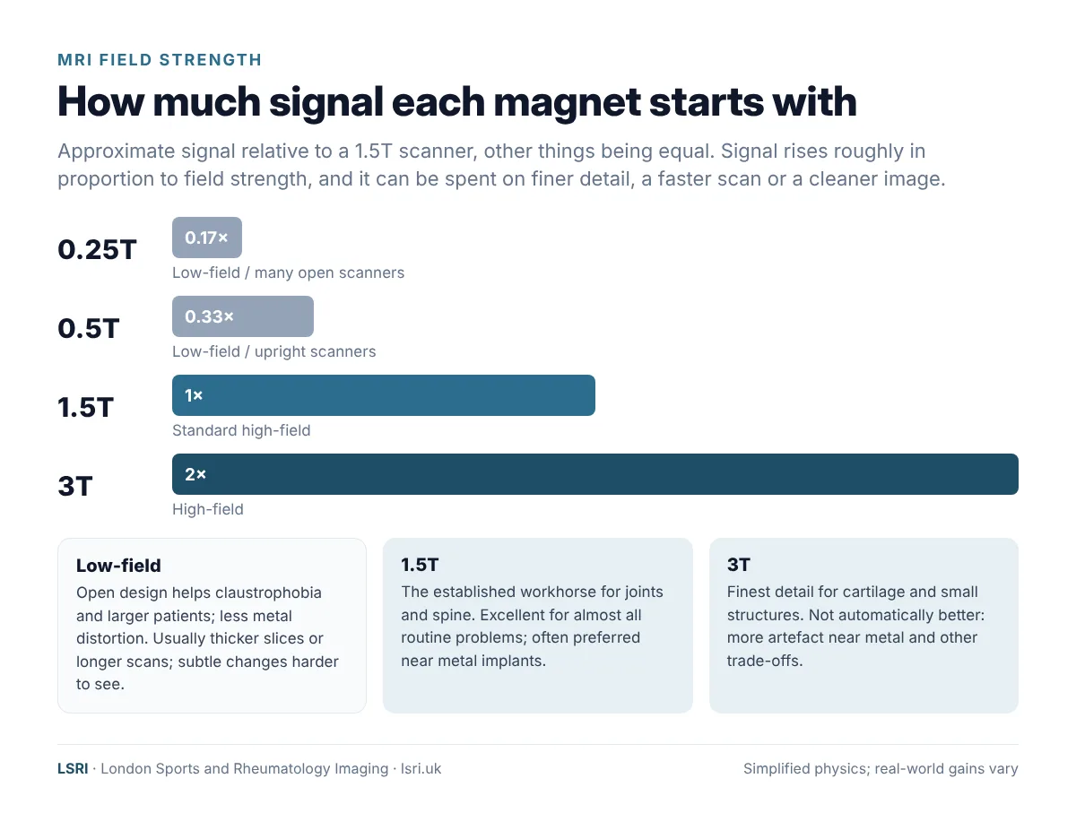
- A stronger magnet gives you more signal to spend. Signal rises roughly in proportion to field strength, so a 3T magnet starts with about twice the signal of a 1.5T magnet, and a 1.5T magnet about six times that of a 0.25T magnet, other things being equal.
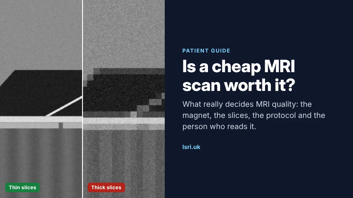
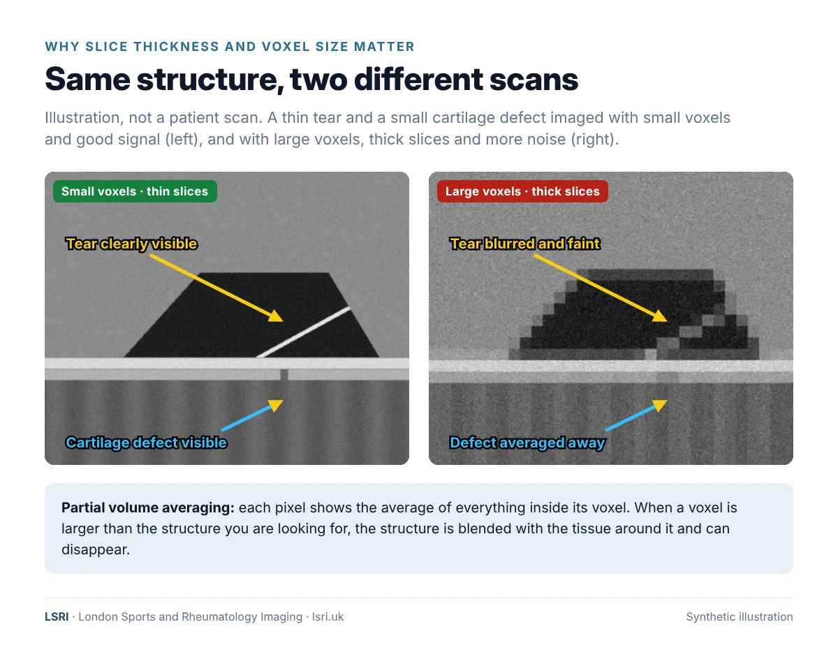
So when a scanner has less signal to work with, or a scan is kept short, something has to give: the slices get thicker, the pixels get bigger, or the image gets grainier. Thick slices matter because of an effect called partial volume averaging. Each pixel shows the average of everything inside its voxel, so a structure thinner than the slice is blended with the tissue around it. A small tear, a thin area of cartilage damage or an early change in the bone marrow can simply be averaged out of sight.
Field strength explained: low-field, 1.5T and 3T
MRI field strength is measured in tesla (T). Clinical scanners in the UK mostly fall into three groups.
| Low-field (about 0.2–0.6T), including many open and upright scanners | 1.5T | 3T | |
|---|---|---|---|
| Signal available | Lowest | Good | About double 1.5T |
| Main strengths | Open design helps with claustrophobia and larger patients; some allow scans standing or sitting; less distortion from metal | The established workhorse for joints and spine; robust; often preferred around metal implants | Finest detail for cartilage and small structures; can scan faster or at higher resolution |
| Main limitations | Lower resolution or longer scans; fat suppression is less effective; subtle cartilage, small tear and marrow changes are harder to see | Slightly less detail than 3T for the very smallest structures | More artefact near metal; more sensitive to some image distortions; stricter limits on tissue heating |
What the research shows about low-field and open MRI
For some large, common problems, low-field and high-field scanners perform similarly. In a study of 114 patients scanned on both a 0.5T and a 1.5T system, accuracy for anterior cruciate ligament (ACL) tears was about the same (90% and 91%).3 A systematic review of knee MRI reached a similar conclusion for meniscal and cruciate ligament tears, but found too little evidence to rely on low-field scanners for cartilage damage and osteoarthritis.4
The differences show up with smaller or subtler problems. In a study of 379 patients scanned on a 0.2T scanner and then examined by keyhole surgery (arthroscopy), the scans identified 85% of ACL tears but only 51% of tears of the lateral meniscus and 8% of osteochondral (cartilage and bone) lesions.5 In the lumbar spine, grading of disc degeneration on a 0.25T scanner agreed only moderately with 3T.6 A 2024 review of modern low-field MRI concluded that image quality is still below 1.5T, scans take longer, and the technique is not suitable for very small structures such as the wrist ligaments, while noting clear advantages around metal and in cost.7
To be fair to the technology, low-field MRI is improving. Small recent studies of new-generation 0.55T scanners using deep-learning (AI) image reconstruction have reported knee results close to 3T.8,9 These are early, small studies, and premium 0.55T systems are not the same as older 0.2–0.3T open scanners. They are a reminder that the label alone does not tell the whole story, which is why it is worth asking the questions further down this page.
Is 3T always better than 1.5T?
Not automatically. 3T delivers about twice the signal of 1.5T, which can be spent on finer detail or a shorter scan.10 In one large comparison, 3T was more accurate than 1.5T for knee cartilage (80% vs 75%).11 But most head-to-head studies in MSK imaging show only a small advantage at best, and some show none: in one study of young patients with knee injuries, 1.5T detected lateral meniscal tears more reliably than 3T.7,12 Stronger magnets also bring more distortion around metal, so 1.5T is generally preferred for people with joint replacements, plates or screws.13,14
The practical message: a modern, well-run 1.5T or 3T scanner with the right coil and protocol is an excellent tool for almost all joint and spine problems. The bigger drops in quality come from low signal, thick slices, rushed protocols and, above all, inexpert reporting.
The parts of a scan you never see: coils, protocols and planning
The coil. The receiver coil is the antenna that picks up the MRI signal. A dedicated coil fitted closely around the knee, shoulder, wrist or ankle collects far more signal from that area than a general-purpose coil, which allows thinner slices without a grainy picture.
The protocol. The protocol is the set of sequences, planes and slice thicknesses used for your scan. A good protocol is tailored to the question: cartilage-sensitive sequences for a suspected cartilage injury, extra planes for the shoulder or hip labrum, intravenous contrast for some lumps, or images of the whole spine when cancer spreading to the spine is a possibility, because spinal metastases are often found at more than one level.15
The planning. The Royal College of Radiologists’ reporting standards make the same point from the other direction: the person requesting the scan should give an accurate history and a clear clinical question, and the report should answer that question directly.16 That is why a short conversation with a radiologist before booking can be worth more than any upgrade to the scanner. It decides which body part is scanned, which sequences are used, whether contrast is needed, and sometimes whether an ultrasound or X-ray would answer the question better and more cheaply. Our guide to MRI or ultrasound for joint pain explains how we choose.
The biggest variable: who reads your scan
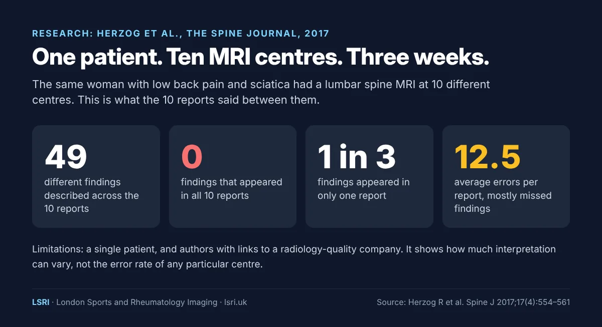
In a study published in The Spine Journal, one 63-year-old woman with low back pain and sciatica had a lumbar spine MRI at 10 different centres within three weeks.17 The 10 reports described 49 different findings between them. Not one finding appeared in all 10 reports, and about a third of the findings appeared in only one. Compared with a reference reading by spine specialists, the reports contained an average of 12.5 errors each, mostly things that were missed. The study involved a single patient, and its authors had links to a company that measures radiology quality, so it should not be over-read. But it shows how much interpretation can vary.
Specialist training makes a measurable difference. In a US study of more than 2,300 second opinions given by MSK subspecialist radiologists, the interpretation changed in a clinically important way in about 1 in 4 cases, and in over a third of tumour cases. Where surgery or a biopsy later confirmed the diagnosis, the second opinion was correct 82% of the time.18 A 2025 review by UK radiologists of 8 studies, covering more than 11,000 examinations, found clinically significant changes in between 1.4% and 27.9% of cases, highest for tumours and lowest for simple limb X-rays.19 In a study of hip MRI checked against arthroscopy, MSK specialists correctly identified cartilage damage in the hip socket 79% of the time, compared with 28% for community radiologists.20
These studies looked at cases sent for a second opinion, which are often more complex than average, so the figures do not apply to every scan. They do show why it is worth knowing who will read yours.
Does it matter where the radiologist is?
Remote reporting (teleradiology) is a normal part of modern radiology, in the NHS and privately. A UK-based specialist reporting from another room, or another city, can be excellent. The question that matters is whether the person reporting your scan works within the UK’s professional framework. The Royal College of Radiologists’ teleradiology standards say the reporting radiologist should be on the GMC Specialist Register for clinical radiology and indemnified to UK standards, should have access to the patient’s clinical details and previous imaging, should work within proper quality assurance, and should be available to discuss the report.21 The College’s 2025 reporting standards add that radiologists reporting remotely for private providers may be unfamiliar with local care pathways, so safeguards need to be in place.16
Why it matters that your radiologist is based in the UK
Some lower-cost scans are sent outside the UK to be reported. Whatever the individual radiologist’s skill, that arrangement can leave you with less protection and a thinner report:
- Accountability. The GMC can only hold to account doctors on its register. If the radiologist who reports your scan is not GMC-registered, there is no UK regulator to turn to if something is missed.
- Indemnity. They may not carry UK-standard medical indemnity, which matters if a missed diagnosis leads to harm.
- Context. A radiologist working through a reporting company may never see your full history or your previous scans, may not know UK referral pathways, and may not be reachable to discuss the findings with you or your clinician.
- Time and care. Remote reporting services, especially if based outside the UK, may not have the full context of your case, and the radiologist may never know who you are or what happened next. A specialist who knows they are answerable for the report, and may be the one explaining it to you, has every reason to take more care.
At LSRI, every scan is reported by a UK-governed consultant musculoskeletal radiologist, on the GMC Specialist Register and indemnified to UK standards, who sees your history and whom you and your clinician can speak to. A scan is only as good as the report that comes with it.
Is a cheaper scan ever the right choice?
Sometimes, maybe, as long as you are told clearly what quality of scan you are getting, who will report it, where that radiologist is based, and whether the provider outsources reporting outside the UK or to another teleradiology company to keep costs down. With that information you can make an informed choice. If you cannot manage a conventional scanner even with support, an open scanner may be the only way to be scanned at all. If the question is simple and the answer is likely to be obvious, a basic scan may answer it. And some problems are better answered by ultrasound or X-ray, which cost less than any MRI. Sometimes no scan is needed at all.
A high-quality scan with specialist reporting matters most when:
- pain is persistent, unexplained or not improving as expected;
- a serious cause such as a fracture, infection or tumour needs to be ruled out;
- the problem involves cartilage, small ligaments, the labrum or the small joints of the hand, wrist or foot;
- the result will decide whether you have an injection or surgery;
- you have already had a scan that did not explain your symptoms.
“Penny wise, pound foolish” is an old saying for a reason. A scan that cannot answer the question often leads to a second scan, more appointments and months of treatment aimed at the wrong problem. Our case studies on nine months of back pain, spinal metastases and four years of shoulder pain show what a delayed diagnosis can mean.
10 questions to ask before you book an MRI
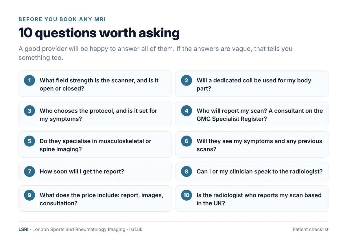
- What is the field strength of the scanner (for example 0.3T, 1.5T or 3T), and is it an open or closed scanner?
- Will a dedicated coil be used for my body part?
- Who chooses the protocol, and is it set for my symptoms?
- Who will report my scan? Are they a consultant radiologist on the GMC Specialist Register?
- Do they specialise in musculoskeletal or spine imaging?
- Will they see my symptoms and any previous scans?
- How soon will I get the report?
- Can I, or my clinician, speak to the radiologist about the findings?
- What does the price include: the report, a copy of my images, a consultation?
- Is the radiologist who reports my scan based in the UK?
How we approach MRI at LSRI
- Advice before you book. Call us and we will help you decide whether MRI, ultrasound or X-ray is the right first test, which area to scan and which protocol to use. If a scan is unlikely to change your treatment, we will say so.
- Specialist reporting. Your MRI is reported by a consultant musculoskeletal radiologist within 24 hours. Our service is led by Dr Prashant Sankaye, Consultant MSK Radiologist.
- Modern scanners. MRI is carried out at our partner imaging centres on 1.5T and 3T scanners, for patients of both our Harley Street and Harrow clinics, and we can arrange scans at 70+ UK locations including partner centres.
- Clear pricing. MRI from £375 per body part, including the scan, a specialist report from a consultant radiologist and access to your images, or from £425 with a consultation. See our pricing page.
- Time to talk. MRI with a consultation, to go through the results and next steps, is from £425.
- Second opinions. Already had a scan elsewhere? We offer second opinions on existing images and reports.
Frequently asked questions
Is 3T MRI better than 1.5T MRI?
For most joint and spine problems, both give excellent results when the scanner, coil and protocol are good. 3T has about twice the signal, which helps with cartilage and very small structures. 1.5T is often preferred if you have metal implants, because metal distorts the image less. The quality of the protocol and the report usually matters more than the difference between the two.
Is an open MRI as good as a closed MRI?
Not usually for detail. Most open and upright scanners are low-field, so they have less signal and usually produce lower-resolution images or need longer scans. They can still answer some questions well, and they are a good option if you cannot tolerate a conventional scanner. If you are anxious about enclosed spaces, ask about a wide-bore scanner, which has a larger opening but keeps high-field image quality.
Why are some private MRI scans so cheap?
Lower prices usually reflect choices about the scanner, the length of the protocol, who reports the scan and what is included afterwards. Those choices are legitimate, but they are trade-offs. Ask the 10 questions above so that you know what you are paying for.
Does it matter if my MRI is reported remotely?
Reporting from another room or another city in the UK can be excellent. What matters is that the radiologist is based in the UK, is a GMC-registered specialist with UK-standard indemnity, sees your symptoms and previous scans, works within proper quality assurance and can be contacted to discuss the findings. If a scan is reported outside the UK, you may have none of those protections, so ask before you book.
Can you give a second opinion on an MRI I have already had?
Yes. Send us your images and report and a consultant MSK radiologist will review them. See our second opinion service, and our guide to understanding MRI scan results.
Do I need a GP referral for a private MRI?
No GP referral is needed in most cases if you are paying for yourself. If you are claiming through private medical insurance, your insurer may require a referral from a GP or specialist. Dr Sankaye is recognised by the major insurers and is Bupa fee-assured, so in some cases your insurer may cover a scan and consultation with him directly. Speak to your insurer first to check what your policy covers.
Book an appointment
Call 0203 633 5040 or book online. Not sure which scan you need? Call us and we will help you choose. Clinics at Harley Street (W1G 9QJ) and Harrow (HA2 7SJ).
References
- Elster AD. Signal-to-noise ratio. Questions and Answers in MRI. mriquestions.com
- Maubon AJ, Ferru JM, Berger V, et al. Effect of field strength on MR images: comparison of the same subject at 0.5, 1.0, and 1.5 T. RadioGraphics 1999;19(4):1057–1067. doi:10.1148/radiographics.19.4.g99jl281057
- Vellet AD, Lee DH, Munk PL, et al. Anterior cruciate ligament tear: prospective evaluation of diagnostic accuracy of middle- and high-field-strength MR imaging at 1.5 and 0.5 T. Radiology 1995;197(3):826–830. doi:10.1148/radiology.197.3.7480763
- Puig S, Kuruvilla YCK, Ebner L, et al. Magnetic resonance tomography of the knee joint. Skeletal Radiology 2015;44(10):1427–1434. doi:10.1007/s00256-015-2178-5
- Lee CS, Davis SM, McGroder C, et al. Analysis of low-field magnetic resonance imaging scanners for evaluation of knee pathology based on arthroscopy. Orthopaedic Journal of Sports Medicine 2013;1(7):2325967113513423. doi:10.1177/2325967113513423
- Hansen BB, Ciochon UM, Trampedach CR, et al. Grading lumbar disc degeneration: a comparison between low- and high-field MRI. Acta Radiologica 2019;60(12):1636–1642. doi:10.1177/0284185119842472
- Pogarell T, Heiss R, Janka R, et al. Modern low-field MRI. Skeletal Radiology 2024;53(9):1751–1760. doi:10.1007/s00256-024-04597-4
- Donners R, Vosshenrich J, Segeroth M, et al. Deep learning reconstructed new-generation 0.55 T MRI of the knee: a prospective comparison with conventional 3 T MRI. Investigative Radiology 2024;59(12):823–830. doi:10.1097/RLI.0000000000001093
- Ulas ST, Hess M, Zhu Z, et al. Diagnostic efficacy of deep learning-based denoising of low-field 0.55 T compared to conventional 3 T knee MRI. European Journal of Radiology 2026;204:113163. doi:10.1016/j.ejrad.2026.113163
- Wong S, Steinbach L, Zhao J, et al. Comparative study of imaging at 3.0 T versus 1.5 T of the knee. Skeletal Radiology 2009;38(8):761–769. doi:10.1007/s00256-009-0683-0
- Kijowski R, Blankenbaker DG, Davis KW, et al. Comparison of 1.5- and 3.0-T MR imaging for evaluating the articular cartilage of the knee joint. Radiology 2009;250(3):839–848. doi:10.1148/radiol.2503080822
- Nouri N, Bouaziz MC, Riahi H, et al. Traumatic meniscus and cruciate ligament tears in young patients: a comparison of 3T versus 1.5T MRI. Journal of the Belgian Society of Radiology 2017;101(1):14. doi:10.5334/jbr-btr.1158
- Jungmann PM, Agten CA, Pfirrmann CW, et al. Advances in MRI around metal. Journal of Magnetic Resonance Imaging 2017;46(4):972–991. doi:10.1002/jmri.25708
- Sneag DB, Abel F, Potter HG, et al. MRI advancements in musculoskeletal clinical and research practice. Radiology 2023;308(2):e230531. doi:10.1148/radiol.230531
- Finucane LM, Downie A, Mercer C, et al. International framework for red flags for potential serious spinal pathologies. Journal of Orthopaedic & Sports Physical Therapy 2020;50(7):350–372. doi:10.2519/jospt.2020.9971
- The Royal College of Radiologists. Standards for interpretation and reporting of imaging investigations, third edition. 2025. rcr.ac.uk
- Herzog R, Elgort DR, Flanders AE, et al. Variability in diagnostic error rates of 10 MRI centers performing lumbar spine MRI examinations on the same patient within a 3-week period. The Spine Journal 2017;17(4):554–561. doi:10.1016/j.spinee.2016.11.009
- Chalian M, Del Grande F, Thakkar RS, et al. Second-opinion subspecialty consultations in musculoskeletal radiology. American Journal of Roentgenology 2016;206(6):1217–1221. doi:10.2214/AJR.15.14540
- Patel A, Isaac A. The clinical value of second-opinion reporting by subspecialist musculoskeletal radiologists. European Journal of Radiology 2025;190:112262. doi:10.1016/j.ejrad.2025.112262
- McGuire CM, MacMahon P, Byrne DP, et al. Diagnostic accuracy of magnetic resonance imaging and magnetic resonance arthrography of the hip is dependent on specialist training of the radiologist. Skeletal Radiology 2012;41(6):659–665. doi:10.1007/s00256-011-1266-4
- The Royal College of Radiologists. Standards for the provision of teleradiology within the United Kingdom, second edition. 2016. rcr.ac.uk
About this article
Written by the LSRI clinical team for patient education, based on the peer-reviewed research and UK professional standards listed above. It is general information, not a diagnosis or a substitute for advice about your own symptoms. LSRI is a private imaging clinic and provides the scans described here; we have tried to present the evidence fairly, including where a cheaper scan, a different test or no scan at all is the right choice. We do not name or comment on other providers.