Written by: LSRI Editorial Team
Medically reviewed by Dr Prashant Sankaye, Consultant MSK Radiologist ·
Last Updated: October 14, 2026
Read Time: 16 Minutes
Written by the LSRI clinical team and medically reviewed by Dr Prashant Sankaye, Consultant MSK Radiologist, on 11 October 2026.
When you book an MRI scan, you usually choose a clinic, a date and a price. The most important choice is often invisible: who will read your scan. An MRI examination produces hundreds of images, but what you take away is the report, and the report is only as good as the person who writes it and the scan they were given to read. This article explains what a musculoskeletal (MSK) radiologist is, what the evidence says about subspecialist reporting, why the scan “protocol” matters, and what second and third opinions are for. It draws on Dr Prashant Sankaye’s experience of reviewing other radiologists’ reports in second opinions, arbitration and complaints.
Key points
- An MSK radiologist is a consultant radiologist who has specialised further in bones, joints, muscles, tendons, ligaments and the spine.
- When MSK specialists review scans first reported elsewhere, clinically significant differences have been found in between 1.4% and 27.9% of cases, highest for tumours and complex cases.1
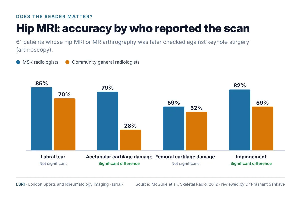
- In one study of hip MRI, MSK radiologists were more accurate than community general radiologists, significantly so for damage to the hip-socket (acetabular) cartilage and for impingement.2
- In knee MRI, the radiologist’s experience mattered more than the strength of the scanner’s magnet.3
- The protocol (how the scan is set up) decides what can be seen. A finding can be invisible if the scan was not designed to show it.4
- When comparing prices, compare what is included: who reports the scan, whether they specialise in MSK imaging and where they are based, how the protocol is chosen, and whether you can talk to the radiologist.
What is an MSK radiologist?
A radiologist is a doctor who specialises in diagnosing illness and injury using imaging. In the UK, consultant radiologists complete years of specialist training, usually pass the Fellowship examinations of the Royal College of Radiologists (FRCR) and are on the General Medical Council’s Specialist Register. Most then focus on one area of the body. A musculoskeletal (MSK) radiologist has specialised in the bones, joints, muscles, tendons, ligaments, nerves and spine, usually with additional fellowship training, and spends most or all of their working week on MSK imaging and image-guided procedures.
All consultant radiologists are highly trained. But radiology now covers so many techniques and conditions that no one can be expert in all of them, and UK reporting standards expect everyone who reports scans to work within a defined scope of practice.5 In busy services, scans are not always matched to a reporter with expertise in that part of the body, and that is where subspecialty experience can make a difference.
Does it matter who reports your MRI? What the evidence shows
Specialists are more accurate in complex areas
In a study of hip MRI and MR arthrography in patients who went on to have keyhole surgery, MSK radiologists and community general radiologists were compared against what the surgeon actually found. The MSK radiologists were more accurate for all four features studied, and significantly so for damage to the cartilage of the hip socket (79% vs 28%) and for impingement (82% vs 59%).2
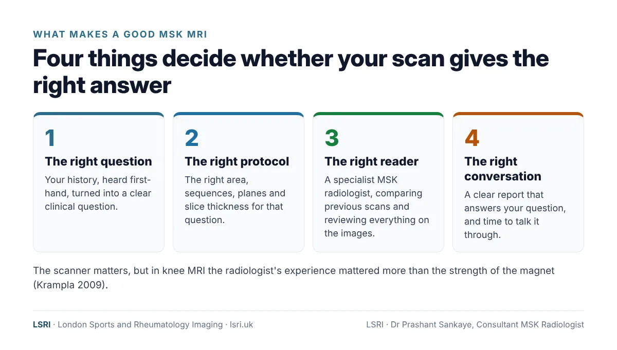
In knee MRI, a study of 11 radiologists found that less experienced readers made significantly more errors for most features, and that the number of errors depended on the reader but not on the strength of the scanner’s magnet. The authors concluded: “Radiologist experience seems to be more important than field strength.”3
Second opinions by MSK specialists find important differences in a minority of cases
Several studies have looked at what happens when MSK radiologists review scans first reported elsewhere:
- At one US academic centre, MSK radiologists reviewed 2,326 outside MRI and CT examinations. 26.2% had clinically important differences, defined as differences likely to change patient management, rising to 36.3% in tumour cases. Among discrepant cases where pathology later confirmed the diagnosis, the second opinion was correct in 82%. Most differences were in interpreting abnormalities rather than detecting them.6
- In orthopaedic oncology (bone and soft-tissue tumours), clinically significant differences were found in 9.2% of scans first read by MSK fellowship-trained radiologists, compared with 27.9% of scans first read by radiologists without MSK training.7
- Across 1,052 MSK second opinions at another US centre, 30% differed from the original report and 18% were judged likely to be clinically relevant.8
- A 2025 systematic review of eight studies and more than 11,000 examinations found clinically significant differences ranging from 1.4% to 27.9%. They were lowest for routine X-rays and knee MRI reports and highest in oncology and complex cross-sectional imaging. The authors concluded that subspecialty MSK interpretations were more accurate than primary interpretations.1
The higher figures come from specialist centres where complex and tumour cases are referred for review, so they are likely to overstate how often a routine report would change. The honest message is that most reports are accurate, but in complex, subtle or high-stakes cases a specialist review changes the picture surprisingly often. The same pattern has been seen in other specialties: in a UK study of specialist neuroradiology second opinions, 13% had major differences that significantly affected patient management.9
Reports on the same patient can vary widely
In one striking study, a 63-year-old woman with back pain and sciatica had lumbar spine MRI at 10 different centres within three weeks. Across the 10 reports there were 49 different findings. Not one was reported by all 10 centres, and a third were reported only once.10 It was a single patient, so it cannot tell us how common such variation is, but it shows clearly that the scan alone does not make the diagnosis.
The protocol: the part of your scan you never see
Before you lie down in the scanner, someone has decided which body part to cover, which sequences to run, in which planes and at what slice thickness, and whether contrast or special techniques are needed. This is the protocol, and it decides what can and cannot be seen.
- It should be tailored to the question. Guidance on knee MRI notes that many practices tailor each study to the specific clinical question, that it is the supervising doctor’s responsibility to decide whether additional or specialised sequences are needed, and that slices in the sagittal and coronal planes should be 4 mm thick or less.4
- Technique can change sensitivity for some questions. For tears of the hip labrum, for example, a meta-analysis found pooled sensitivity of about 80% for MRI overall and 89% for MR arthrography, with 3 tesla MRI close to arthrography, so the technique can affect how many tears are found.11
- Some conditions have specific requirements. European guidelines for soft-tissue tumours recommend a scanner of at least 1.5 tesla, high-resolution axial images to show the tumour’s margins, and a skin marker over the lump, and advise that suspected sarcomas are referred to a specialist tumour centre before biopsy or surgery.12
- A bigger magnet is not the whole story. For ACL and meniscal tears of the knee, a meta-analysis found that magnetic field strength had no significant effect on accuracy.13 The protocol and the reader matter at least as much as the machine.
In UK hospital departments, routine requests are usually vetted and protocolled by experienced radiographers, with complex or specialist examinations more likely to be vetted by a radiologist; the guidance says both should be able to add or edit protocols.14 At LSRI, protocols are planned around your symptoms and your question, by a team led by a consultant MSK radiologist.
What makes a good MSK MRI report?
A good report is more than a list of observations. It should:
- answer the clinical question that was asked, clearly and early;
- describe what is relevant to your symptoms, and say which findings are probably incidental or age-related;
- compare with previous imaging, or say if previous imaging was not available and that matters;5
- grade severity where it affects treatment (for example, a partial or complete tear, or a low- or high-risk stress fracture);
- mention any limitations, such as movement on the images or a missing sequence;
- end with a clear conclusion and, where appropriate, a recommendation for the next step.
UK standards also expect clinical details to form part of the report, and say that interpretation may be incorrect if referral details are deficient, absent or misleading.5 That is why, at LSRI, the radiologist talks to you. See the radiology consultation: why we talk to you first.
The UK picture: workforce pressures and remote reporting
Radiology in the UK is short of specialists. The Royal College of Radiologists’ 2025 census found a 32% shortfall of consultant radiologists, and NHS spending across the UK on outsourcing, insourcing and locums reached £362 million in 2025. Outsourcing alone was over £241 million, covering the reporting of around 28% of CT and 24% of MRI scans.15
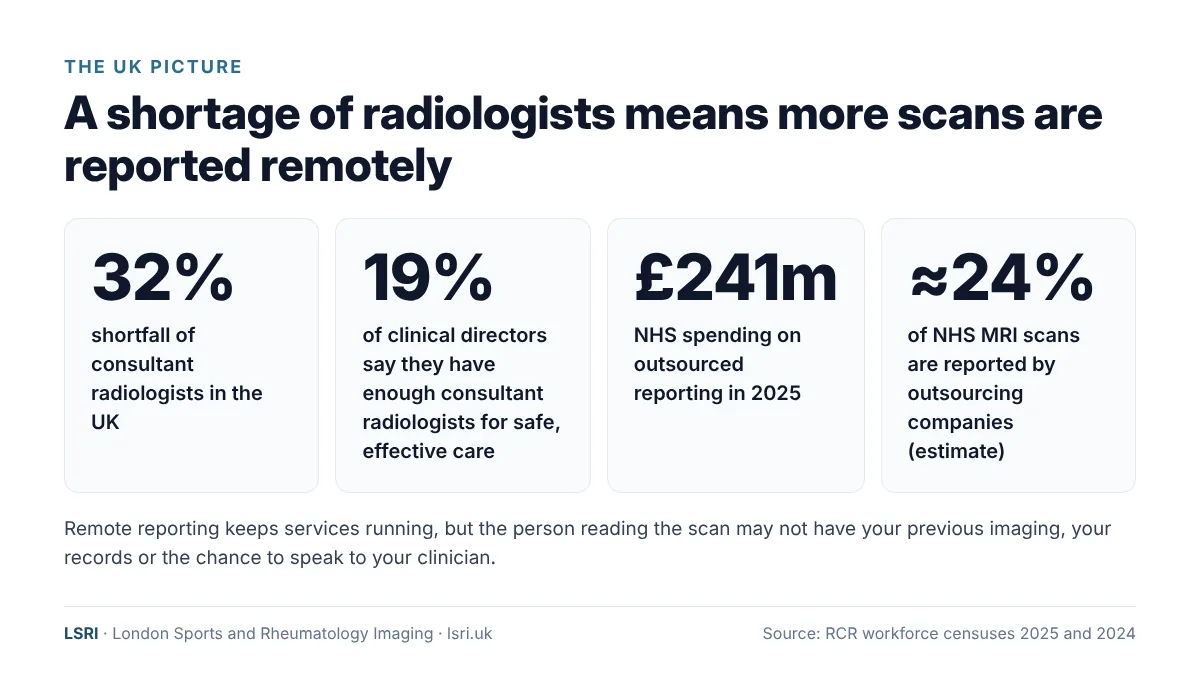
The Royal College of Radiologists has also noted that teleradiology reporters often lack access to patients’ previous imaging and electronic records, and may not be able to speak to the clinician looking after the patient.16 Its standards say teleradiologists must have access to the same breadth of patient information, including previous images and reports, as they would have in the hospital itself.17 Many radiologists who report remotely are skilled specialists, and outsourcing keeps services running. But remote reporting services, especially if based outside the UK, may not have the full context of your case, and the radiologist may never know who you are or what happened next.
What are you paying for?
It is natural to compare scans on price. It is also worth comparing what the price includes, because the scan is only half of what you are buying; the other half is the expertise that turns images into a diagnosis, and the report is what guides your treatment. Our case studies on a hip stress fracture and a heel (calcaneal) fracture found on MRI after eight months of pain show how much an accurate diagnosis matters. Before you book, ask:
- Who will report my scan, and do they specialise in musculoskeletal imaging?
- Is the radiologist who reports my scan based in the UK?
- How is the scan protocol chosen for my problem?
- Will the radiologist see my history and any previous scans?
- Can I talk to the radiologist about the result?
For more on this, see Is a cheap MRI scan worth it?
Second and tertiary opinions: what they are for
- A second opinion is an independent review of your images, usually by a radiologist with specialist expertise in that area, to confirm or refine the original report.
- A tertiary (third) opinion is sought when two opinions disagree, or when a case is complex enough to need a further specialist view.
- Arbitration is an independent decision on a disputed report, for example when a reporting provider and the clinician or organisation that commissioned the report disagree about whether something was missed.
Dr Prashant Sankaye has provided second and tertiary opinions, and independent arbitration in disputes about radiology reports. As a former Head of Specialty at Imperial College Healthcare NHS Trust, his responsibilities included handling complaints and reviewing reporting discrepancies, and as a former Expert Advisor to the Parliamentary and Health Service Ombudsman he gave expert radiology advice on complaints about NHS care.
What claims data show
Problems with diagnosis are the most common reason for claims involving radiology. In the United States, errors in diagnosis are by far the most common cause of malpractice claims against radiologists, and fractures are among the diagnoses most often missed.18 In England, of 1,472 radiology claims closed with damages by NHS Resolution between 2019/20 and 2023/24, around two-thirds (961) had a primary cause of failure to interpret an X-ray, failure or delay in diagnosis, or wrong diagnosis.19
Claims are rare compared with the very large number of scans performed each year. In Dr Sankaye’s experience of reviewing complaints and disputes, the same themes come up again and again: missing clinical information, previous scans not compared, the wrong area or protocol, reading outside one’s usual field, and reports that do not answer the question asked.
“Reviewing difficult cases, through second opinions and complaints, has shown me how easily things can be missed, and it shapes how I work. No radiologist can promise never to miss anything. But taking time to understand the patient, choosing the right protocol and reading as a specialist all help.”
Dr Prashant Sankaye, Consultant MSK Radiologist
If you are a clinician, solicitor or insurer needing an independent expert opinion on musculoskeletal imaging, see our medicolegal services. For patients, see our MRI second opinion service.
How LSRI can help
- Specialist reporting. Every LSRI report is written by a UK-governed consultant musculoskeletal radiologist, usually within 24 hours.
- Clear pricing. MRI from £375 per body part, including the scan, a specialist report and access to your images.
- Time to talk. MRI with a consultation, to go through the results and next steps, is from £425.
- Independent second opinions on MRI, CT, X-ray and ultrasound performed elsewhere.
Read more: Can an MRI miss something? and about Dr Prashant Sankaye.
Frequently asked questions
What is the difference between a radiologist and an MSK radiologist?
All radiologists are doctors trained to interpret medical images. An MSK radiologist has specialised further in bones, joints, muscles, tendons, ligaments and the spine, and spends most of their time on this work, including image-guided procedures such as ultrasound-guided injections.
How do I find out who will report my scan?
Ask the clinic before you book: it is reasonable to ask who will report your scan, whether they specialise in musculoskeletal imaging and where they are based. The report itself should state the name of the radiologist who wrote it.5
Does a 3 tesla scanner mean a better result?
Not on its own. Higher field strength can help for some questions, but for ACL and meniscal tears a meta-analysis found no significant effect of field strength on accuracy,13 and in another study the reader’s experience mattered more than the magnet.3
Can you review a scan done at another clinic or in the NHS?
Yes. We can review MRI, CT, X-ray and ultrasound images from other providers, ideally with the original report and any previous scans. See our second opinion page for how to send them.
What happens if the second opinion disagrees with my original report?
We explain what we found and why, in writing and in a consultation, and, with your permission, share our report with your clinician so that they can decide on the next steps with you.
Book an appointment
Call 0203 633 5040 or book online. Clinics at Harley Street (W1G 9QJ) and Harrow (HA2 7SJ). If you have emergency symptoms, go to A&E or call 999 instead.
References
- Patel A, Isaac A. The clinical value of second-opinion reporting by subspecialist musculoskeletal radiologists. European Journal of Radiology 2025;190:112262. doi:10.1016/j.ejrad.2025.112262
- McGuire CM, MacMahon P, Byrne DP, et al. Diagnostic accuracy of magnetic resonance imaging and magnetic resonance arthrography of the hip is dependent on specialist training of the radiologist. Skeletal Radiology 2012;41(6):659–665. doi:10.1007/s00256-011-1266-4
- Krampla W, Roesel M, Svoboda K, et al. MRI of the knee: how do field strength and radiologist’s experience influence diagnostic accuracy and interobserver correlation in assessing chondral and meniscal lesions and the integrity of the anterior cruciate ligament? European Radiology 2009;19(6):1519–1528. doi:10.1007/s00330-009-1298-5
- American College of Radiology, Society for Pediatric Radiology, Society of Skeletal Radiology. ACR–SPR–SSR practice parameter for the performance and interpretation of magnetic resonance imaging (MRI) of the knee. Revised 2025. acr.org
- The Royal College of Radiologists. Standards for interpretation and reporting of imaging investigations, third edition. October 2025. rcr.ac.uk
- Chalian M, Del Grande F, Thakkar RS, et al. Second-opinion subspecialty consultations in musculoskeletal radiology. American Journal of Roentgenology 2016;206(6):1217–1221. doi:10.2214/AJR.15.14540
- Rozenberg A, Kenneally BE, Abraham JA, et al. Second opinions in orthopedic oncology imaging: can fellowship training reduce clinically significant discrepancies? Skeletal Radiology 2019;48(1):143–147. doi:10.1007/s00256-018-3024-3
- Bedoya MA, Chi AS, Harsha AK, et al. Musculoskeletal Outside Interpretation (MOI-RADS): an automated quality assurance tool to prospectively track discrepancies in second-opinion interpretations in musculoskeletal imaging. Skeletal Radiology 2021;50(4):723–730. doi:10.1007/s00256-020-03601-x
- Briggs GM, Flynn PA, Worthington M, et al. The role of specialist neuroradiology second opinion reporting: is there added value? Clinical Radiology 2008;63(7):791–795. doi:10.1016/j.crad.2007.12.002
- Herzog RJ, Elgort DR, Flanders AE, Moley PJ. Variability in diagnostic error rates of 10 MRI centers performing lumbar spine MRI examinations on the same patient within a 3-week period. The Spine Journal 2017;17(4):554–561. doi:10.1016/j.spinee.2016.11.009
- Zhang P, Li C, Wang W, et al. 3.0 T MRI is more recommended to detect acetabular labral tears than MR arthrography: an updated meta-analysis of diagnostic accuracy. Journal of Orthopaedic Surgery and Research 2022;17(1):126. doi:10.1186/s13018-022-02981-1
- Noebauer-Huhmann IM, Vanhoenacker FM, Vilanova JC, et al. Soft tissue tumor imaging in adults: European Society of Musculoskeletal Radiology–Guidelines 2023—overview, and primary local imaging: how and where? European Radiology 2024;34(7):4427–4437. doi:10.1007/s00330-023-10425-5
- Phelan N, Rowland P, Galvin R, O’Byrne JM. A systematic review and meta-analysis of the diagnostic accuracy of MRI for suspected ACL and meniscal tears of the knee. Knee Surgery, Sports Traumatology, Arthroscopy 2016;24(5):1525–1539. doi:10.1007/s00167-015-3861-8
- The Royal College of Radiologists. Vetting (triaging) and cancellation of inappropriate radiology requests. May 2021. rcr.ac.uk
- The Royal College of Radiologists. Clinical radiology workforce census 2025. 2026. rcr.ac.uk
- The Royal College of Radiologists. Clinical radiology workforce census 2024. 2025. rcr.ac.uk
- The Royal College of Radiologists. Standards for the provision of teleradiology within the United Kingdom, second edition. December 2016. rcr.ac.uk
- Whang JS, Baker SR, Patel R, et al. The causes of medical malpractice suits against radiologists in the United States. Radiology 2013;266(2):548–554. doi:10.1148/radiol.12111119
- NHS Resolution. Freedom of Information request FOI_6728: radiology claims. September 2024. resolution.nhs.uk
About this article
Written by the LSRI clinical team to help patients understand how the choice of radiologist and scan protocol affects their diagnosis, based on the research and guidance listed above. It is general information, not a diagnosis. Nothing in this article is a criticism of any individual radiologist, hospital, teleradiology company or reporting provider. Dr Sankaye’s arbitration, complaints and second-opinion work is described in general terms only; no case, patient or organisation is identified.