Written by: LSRI Editorial Team
Medically reviewed by Dr Prashant Sankaye, Consultant MSK Radiologist ·
Last Updated: October 24, 2026
Read Time: 23 Minutes
Written by the LSRI clinical team and medically reviewed by Dr Prashant Sankaye, Consultant MSK Radiologist, on 24 October 2026.
If you have looked at your MRI images or read your report, you may have seen labels such as T1, T2, PD FS or STIR, and numbers such as “3 mm”. What is the difference between T1 vs T2 MRI, and why should it matter to you? These labels describe how your scan was set up, and the set-up decides what the scan can and cannot show.
Most MRI scans are done well. But two scans of the same knee or hip can be very different, depending on the scanner, the coil, the sequences chosen, how thin the slices are and how long the scan was allowed to take. A scan that was not set up to answer your question can look “normal” and still miss the problem. That can falsely reassure you and delay the right treatment. This guide, written by the LSRI clinical team, led by a consultant musculoskeletal (MSK) radiologist, explains what the labels mean, what a well-planned scan involves, and what to ask before you book.
Key points
- An MRI scan is a set of sequences, not one picture. On T1 images fat is bright and fluid is dark; on T2 and fat-suppressed images fluid is bright. Each shows different problems.1,2
- Different questions need different sequences and planes. A protocol that suits one question can miss another, so the scan should be planned for your symptoms.3,4
- Thin slices matter for small structures. Guidelines for the knee and shoulder call for slices of 4 mm or less; small tears can be missed on thicker slices.1,5,6
- The scanner, the coil and the scan time all trade off against detail. A stronger magnet cannot always make up for the wrong coil or the wrong plan.7,8
- Who reads the scan matters too. In one large study of scans sent for a second opinion, specialist MSK radiologists found clinically important differences in 26%.9
- A cheaper scan is not automatically a poor one, but the price can hide real differences in the scanner, the sequences, the slice thickness, the time allowed and who reports it. Ask before you book: see “Cheaper scans and booking-only services” below.
- A “normal” report from a scan that could not show the problem is not the same as a normal joint. If your symptoms do not fit the result, ask for the scan to be reviewed.
How MRI makes a picture, in brief
MRI uses a strong magnet and radio waves to pick up signals from the hydrogen in water and fat in your body. By changing the timing of the radio pulses, the scanner can make different tissues look bright or dark. Each set of settings is called a sequence, and each sequence is repeated as a stack of thin slices through the body part. A typical joint scan uses four to six sequences, usually in all three directions (planes), and the whole set is called the protocol.3,10
Two timings matter most. The repetition time (TR) is how often the radio pulse is repeated. The echo time (TE) is how long the scanner waits before it listens for the signal. Short TR and short TE give a T1-weighted image. Long TR with a moderate-to-long TE gives a T2-weighted, fluid-sensitive image. Long TR with a short TE gives a proton density (PD) image.1,10
T1 vs T2 MRI and the other sequences: what the labels mean
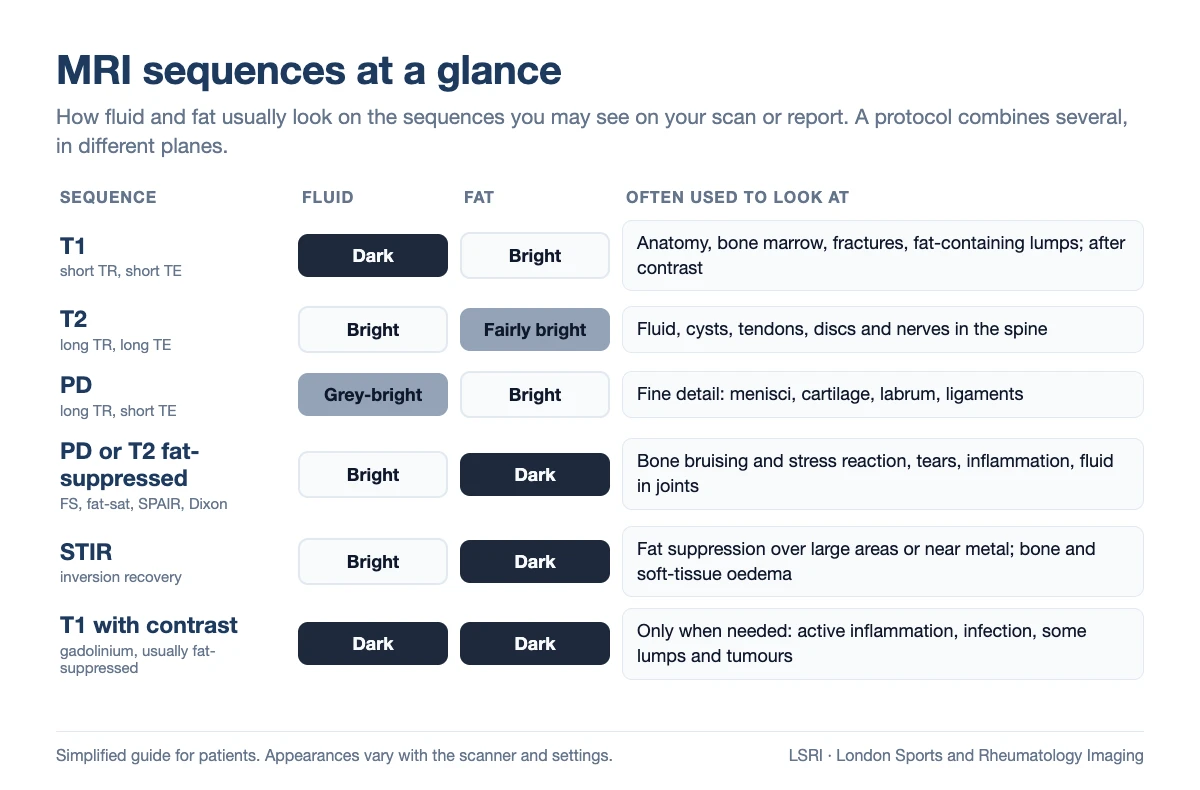
- T1. Fat is bright and fluid is dark, so T1 images show anatomy clearly. They are especially useful for bone marrow: fractures, marrow changes and fat-containing lumps. European guidance says T1 images are still essential in knee MRI.3,5
- T2. Fluid is bright. T2 images show fluid, cysts, swelling and many tendon and spine problems. Because they use a longer echo time, they are less affected by the “magic angle” effect described below.1,11
- PD (proton density). High-detail images, often used for the menisci, cartilage, labrum and ligaments.10
- Fat-suppressed (FS, fat-sat, SPAIR or Dixon). A PD or T2 sequence with the signal from fat suppressed (turned down), so that fluid and swelling stand out. These are the workhorse images for bone bruising, stress reactions, tears and inflammation.2 European knee guidance uses fat-suppressed images in all three planes.10
- STIR. Another way of suppressing fat. It gives even fat suppression over large areas and near metal, but with less signal, so images are a little grainier.1,2
- T1 with contrast (gadolinium). Used only when the question needs it, for example for some infections, active inflammation or lumps. Most joint scans do not need contrast. MR arthrography, where contrast is injected into the joint first, is sometimes used for the hip or shoulder labrum.12,13
One pitfall shows why the choice of sequence matters. A tendon lying at about 55 degrees to the scanner’s magnetic field can look bright on short-echo-time images (T1, PD and gradient echo), as if it were damaged. This is called the magic angle effect. Images with a longer echo time make it disappear, so a protocol needs the right mix of sequences to tell a real tendon problem from an artefact.11
Planes: why one direction is not enough
Slices can be taken from the side (sagittal), from the front (coronal) or across the body part (axial), and sometimes along an angle that follows the anatomy, such as the line of a shoulder tendon or the neck of the femur. Unless a 3D sequence is used, the knee should be scanned in three planes.3 Some structures are only seen well in one direction. A thin structure that looks blurred in one direction, because it runs at an angle through the slices, can be seen clearly in another.
3D sequences, made of tiny cube-shaped blocks of data, can be rebuilt in any direction after the scan. This is part of post-processing. It is useful, and in most centres it is used alongside, rather than instead of, standard 2D sequences.
Slice thickness and gaps: where small problems hide
MRI builds up a body part from a stack of slices. Each slice shows an average of everything within its thickness, and there may be a small gap between slices. A small tear can fall into a gap, or be averaged with the normal tissue around it and fade from view. Radiologists call this partial volume averaging.6
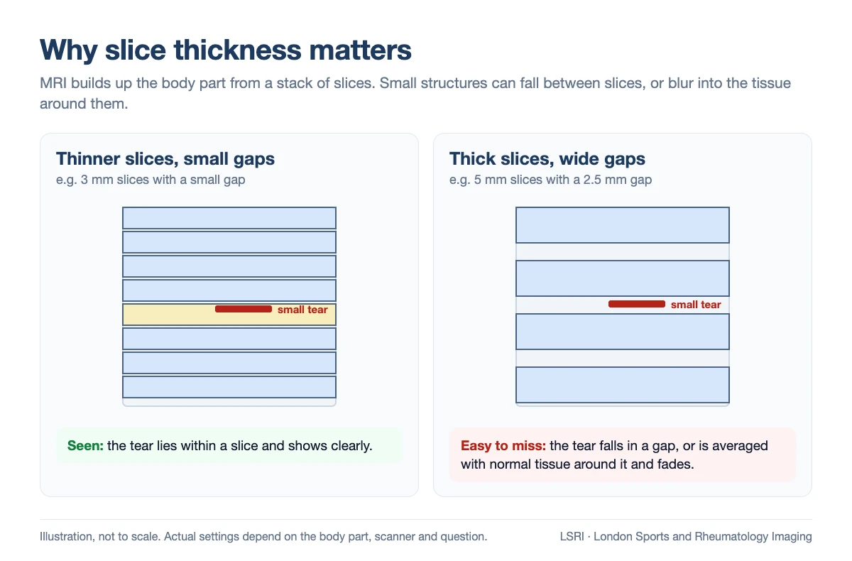
For this reason, guidelines set limits:
- Knee: slices of 4 mm or less in the sagittal and coronal planes “to demonstrate subtle meniscal pathology”, a field of view (the area covered by each image) of 16 cm or less, and a coil placed around the knee.5 European guidance adds that 3 mm or less is preferable and available on most modern scanners.3
- Shoulder: slices of 4 mm or less in the main planes, with a field of view of 16 cm or less.1
- Spine: slices of 4 mm or less in the lower back (3 mm or less in the neck), and gaps between slices are generally not recommended.14
- Hip: 6–8 mm slices are adequate only for screening the whole pelvis. Detailed questions about the hip joint itself, such as the labrum or cartilage, need thinner slices and a small field of view centred on one hip.4,12
Thinner slices are not free. They give less signal in each slice, so they need a stronger magnet, a better coil, more time, or a combination of these.6
Scanner, coil and time: the three-way trade-off
Every MRI scan balances detail against noise and time. The scanner’s field strength, measured in tesla (T), sets how much signal there is to work with: a 3T scanner gives roughly twice the signal-to-noise ratio (the amount of useful signal compared with background noise) of a 1.5T scanner.8 Low-field and many “open” scanners give less signal, so they need longer scans or accept lower resolution.15,16
- Stronger is not automatically better. At 3T, metal implants and some tissues cause more artefacts, so 1.5T is often preferred for people with metalwork.5,8 For knee menisci and ligaments, studies comparing 1.5T and 3T found no significant difference in accuracy, while 3T did better for cartilage.17,18,19
- Low field can still work for some questions. A 2024 review summarised studies in which low-field scanners performed well for some knee injuries, but one shoulder study at 0.2T picked up only 20% of SLAP tears (tears of the labrum at the top of the shoulder socket).16
- The coil matters too. The coil is the part placed around the body part to pick up the signal. US guidelines say a coil placed around the knee (a local coil) is mandatory.5 In one small study of 17 patients, a 3T scanner using its general-purpose body coil gave visibly less signal than a 1.5T scanner with a dedicated knee coil, and picked up 30 of the 33 knee lesions.7
- Time is part of quality. Shorter protocols save scanner time. In one multi-centre study, a well-designed 5-minute knee protocol matched the standard protocol for menisci and ligaments, but was slightly less sensitive for cartilage defects.20 Very short time slots can also mean fewer sequences, fewer planes or thicker slices.
- Movement blurs images. In one US study, 7.5% of outpatient scans had significant motion artefact, and at least one sequence was repeated in about one in five scans.21 Comfortable positioning, padding and time to repeat a blurred sequence all help.
Before and after the scan: planning and processing
Much of what makes a good scan happens before the first image is taken:
- Protocolling: choosing the sequences, planes and coverage for your question, ideally by a radiologist who has read your symptoms.
- Safety screening and positioning: checking for implants, then placing the right coil snugly around the body part so that it stays still.
- Planning the slices: lining them up with the anatomy, for example along a tendon or the femoral neck, and making sure they cover the area that hurts.
After the scan, images can be processed further: 3D data can be rebuilt in other planes, and many new scanners use artificial intelligence (deep learning) to clean up images taken faster. Used well, this can work: in one trial, deep-learning reconstruction nearly halved knee scan time and gave equivalent diagnoses.22 But it has limits. In an international research challenge, a meniscal tear was not visible on images that had been accelerated too far, and standard image-quality scores did not fully match how radiologists ranked the images.23 A clean-looking image is not always a complete one.
Examples: when a scan is not set up for the question
The settings matter most for small structures and early changes. Two common examples:
Medial plica of the knee
A plica is a normal fold in the lining of the knee. In some people, the fold on the inner side of the kneecap thickens and causes pain (plica syndrome). On MRI it shows up most clearly as a dark band outlined by joint fluid, on fluid-sensitive images, especially across the knee (axial).24
In one study, axial images alone detected about three in four symptomatic plicae, and combining two planes raised this to 95%, at the cost of more false alarms.25 A recent study found MRI reliable for detecting a plica but much less reliable for grading it.26 Plicae are also common: in one study of knees that went on to keyhole surgery, MRI showed a plica in about 70%, and its appearance did not predict which were removed. So the finding has to fit your examination and story.27
Hip pain from a labral tear or impingement
A scan of the whole pelvis with thick slices, on its own, is a different test from a dedicated hip MRI with thin slices and a small field of view. For suspected impingement, expert consensus recommends a small field of view on one hip, with radial images (slices arranged like the spokes of a wheel around the hip socket), alongside a fluid-sensitive sequence of the whole pelvis.4
In meta-analyses, standard MRI picked up about two-thirds of labral tears in older studies, and newer 3T scans about 87%, similar to MR arthrography.13,28 The radiologist matters too: in one hip study, specialist MSK radiologists were accurate for damage to the cartilage of the hip socket in 79% of cases, compared with 28% for radiologists without specialist MSK training.29
In both examples, a scan that was not designed for the question can be reported as normal while the cause of your pain is still there.
Why a poor scan can falsely reassure you
A missed finding is not just a technical problem. Missed or delayed diagnoses can lead to harm and missed chances for treatment.30 When a report says “normal”, people often stop looking for an answer, may be told their pain is not structural, or start treatment aimed at the wrong problem. A scan that has to be repeated, or that delays the right treatment, can end up costing more than it saved.
Errors in radiology have several causes: most often a finding is not seen, but the protocol used also affects what can be detected.30 Who reads the scan matters too. In a large US study of CT and MRI scans sent to one academic centre for a second opinion by MSK specialist radiologists, 26% had clinically important differences from the original report; where the diagnosis was later confirmed, the second opinion was correct in 82%.9
In a knee study with several readers and three scanner strengths, errors depended more on the radiologist than on the scanner.31 Reports can also vary between imaging centres scanning the same patient.32 And in most studies, radiologists interpreted scans more accurately when they knew the patient’s clinical history.33
The main UK guidance on MRI equipment, from the medicines and devices regulator (MHRA), focuses on safety rather than on how each scan should be set up for a particular question.34 The Royal College of Radiologists and the College of Radiographers run a voluntary quality scheme for imaging services, the Quality Standard for Imaging (QSI), which includes equipment quality checks, protocols and reporting review.35 So it is reasonable to ask questions.
Cheaper scans and booking-only services: what can differ
A low price does not mean a poor scan, and an expensive one is not automatically better. But when scans are sold mainly on price, these are the things that can differ:
- the scanner’s field strength, and whether a dedicated coil is used for your body part;
- how many sequences and planes are taken, how thin the slices are, and how long the scan slot is;
- whether anyone checks the protocol against your symptoms, or a standard protocol is run for everyone;
- who reports the scan, whether they specialise in muscles, bones and joints, and whether they know your story;
- whether you can speak to anyone about the result.
Resellers (booking-only services) arrange scans at imaging centres run by other companies. Many clinics, including ours, also use partner imaging centres, and booking services can be quick and convenient. Whoever you book with, it is worth asking how your scan will be set up and who will report it. Our guides on whether a cheap MRI scan is worth it and what to check before you book a private MRI online cover price and booking in more detail.
Six questions to ask about the scan itself
- What strength is the scanner, and will a dedicated coil be used for my body part?
- Which sequences and planes are planned, and does someone check them against my symptoms?
- How thin are the slices? For most joints, guidelines suggest about 4 mm or less.1,5
- How long is the scan slot?
- Who reports the scan, do they specialise in MSK imaging, and can I speak to them?
- Will I get my images, so that they can be reviewed later if needed?
If you already have a scan, the report often has a “technique” section listing the sequences, and the image viewer usually labels each series (for example “SAG PD FS” or “COR T1”) and shows the slice thickness. This tells you what was done, but not whether it was right for your question: images need expert interpretation alongside your symptoms.
How we approach MRI at LSRI
LSRI is a doctor-led musculoskeletal imaging clinic. MRI scans are done at partner imaging centres in London and across the UK, and our own clinical team looks after your request and your report.
- Your request is reviewed by a consultant MSK radiologist, who checks that MRI is the right test and which body part to scan.
- Where possible, and when your question needs it, the scan protocol can be adjusted to your symptoms. Many scans are answered well by the standard protocol for that body part.
- Scans at our partner imaging centres are usually on 1.5T scanners, and 3T scanners are also available in London and Nottingham.
- Your scan is reported by a consultant MSK radiologist on the GMC Specialist Register, usually within 24 hours, with your clinical details, and you get your images.
- Two clear MRI options. Scan only, from £375 per body part, includes the scan, the report and your images, but not a consultation or advice on treatment. Scan with a consultation, from £425, adds a consultation with a consultant MSK radiologist to go through your images, the results and next steps.
- A second opinion on a scan you have already had elsewhere: we look at your images and report, answer as much of your question as the available sequences allow and, if needed, advise a repeat scan or a different test, such as an ultrasound.
Our clinics are on Harley Street (W1G 9QJ) and in Harrow (HA2 7SJ); call 0203 633 5040. For more on why the reader matters, see who reports your MRI matters and can an MRI miss something?
Frequently asked questions
What is the difference between T1 and T2 MRI?
On T1 images fat is bright and fluid is dark, which shows anatomy and bone marrow clearly. On T2 images fluid is bright, which shows swelling, cysts and many tendon and spine problems. Most scans use both, often with fat-suppressed versions, because each shows different things.
What does PD fat sat mean on an MRI?
PD stands for proton density, a high-detail sequence often used for cartilage, menisci and ligaments. “Fat sat” (fat saturation, or FS) means the signal from fat has been suppressed, so that fluid, swelling and tears stand out. PD fat-sat images are a mainstay of joint MRI.
What is a STIR sequence?
STIR (short tau inversion recovery) is another way of suppressing the signal from fat, so that fluid and swelling look bright. It works evenly over large areas and near metal, but the images are a little grainier than other fat-suppressed sequences.
What slice thickness should an MRI have?
It depends on the body part and the question. For the knee and shoulder, US guidelines call for slices of 4 mm or less, and European knee guidance prefers 3 mm or less. For the spine, US guidance suggests 4 mm or less, and 3 mm or less for the neck. Thicker slices are adequate for screening a large area such as the whole pelvis, but not for small structures such as the hip labrum.
Can a poor-quality MRI miss a tear?
Yes. Small tears can be missed if the slices are too thick, the wrong sequences or planes are used, the image is blurred by movement, or the scan is accelerated too far. They can also be missed when the scan is read without knowing your symptoms. If your symptoms do not fit a normal result, ask for the scan to be reviewed.
Can you review an MRI scan I had somewhere else?
Yes. As part of a second opinion, a consultant MSK radiologist looks at your images and report and answers as much of your question as the available sequences allow. If needed, we will advise whether a repeat scan or a different test, such as an ultrasound, would help.
References
- American College of Radiology, Society for Pediatric Radiology, Society of Skeletal Radiology. ACR–SPR–SSR practice parameter for the performance and interpretation of magnetic resonance imaging (MRI) of the shoulder. Revised 2024 (Resolution 18). (accessed 24 October 2026) gravitas.acr.org
- Del Grande F, Santini F, Herzka DA, Aro MR, Dean CW, Gold GE, et al. Fat-suppression techniques for 3-T MR imaging of the musculoskeletal system. Radiographics 2014;34(1):217-33. doi:10.1148/rg.341135130
- Parkar AP, Adriaensen MEAPM. ESR essentials: MRI of the knee—practice recommendations by ESSR. Eur Radiol 2024;34:6590-9. doi:10.1007/s00330-024-10706-7
- Castro MO, Mascarenhas VV, Afonso PD, et al. The Lisbon Agreement on femoroacetabular impingement imaging—part 3: imaging techniques. Eur Radiol 2021;31(7):4652-68. doi:10.1007/s00330-020-07501-5
- American College of Radiology, Society for Pediatric Radiology, Society of Skeletal Radiology. ACR–SPR–SSR practice parameter for the performance and interpretation of magnetic resonance imaging (MRI) of the knee. Revised 2025 (Resolution 7). (accessed 24 October 2026) gravitas.acr.org
- Bolog NV, Andreisek G. Reporting knee meniscal tears: technical aspects, typical pitfalls and how to avoid them. Insights Imaging 2016;7(3):385-98. doi:10.1007/s13244-016-0472-y
- Lutterbey G, Behrends K, von Falkenhausen M, Wattjes MP, Morakkabati N, Gieseke J, et al. Is the body-coil at 3 Tesla feasible for the MRI evaluation of the painful knee? A comparative study. Eur Radiol 2007;17(2):503-8. doi:10.1007/s00330-006-0219-0
- Khodarahmi I, Fritz J. The value of 3 Tesla field strength for musculoskeletal magnetic resonance imaging. Invest Radiol 2021;56:749-63. doi:10.1097/RLI.0000000000000801
- Chalian M, Del Grande F, Thakkar RS, Jalali SF, Chhabra A, Carrino JA. Second-opinion subspecialty consultations in musculoskeletal radiology. AJR Am J Roentgenol 2016;206(6):1217-21. doi:10.2214/AJR.15.14540
- European Society of Skeletal Radiology (ESSR) Sports Subcommittee. Guidelines for MR imaging of sports injuries: knee. 2016. (accessed 24 October 2026) essr.org
- Peh WCG, Chan JHM. The magic angle phenomenon in tendons: effect of varying the MR echo time. Br J Radiol 1998;71(841):31-6. doi:10.1259/bjr.71.841.9534696
- American College of Radiology, Society for Pediatric Radiology, Society of Skeletal Radiology. ACR–SPR–SSR practice parameter for the performance and interpretation of magnetic resonance imaging (MRI) of the hip and pelvis for musculoskeletal disorders. Revised 2026 (Resolution 8). (accessed 24 October 2026) gravitas.acr.org
- Smith TO, Hilton G, Toms AP, Donell ST, Hing CB. The diagnostic accuracy of acetabular labral tears using magnetic resonance imaging and magnetic resonance arthrography: a meta-analysis. Eur Radiol 2011;21:863-74. doi:10.1007/s00330-010-1956-7
- American College of Radiology, American Society of Neuroradiology, Society for Advanced Body Imaging, Society of Skeletal Radiology. ACR–ASNR–SABI–SSR practice parameter for the performance of magnetic resonance imaging (MRI) of the adult spine. Revised 2023 (Resolution 6). (accessed 24 October 2026) gravitas.acr.org
- Kravchenko D, Hagar MT, Vecsey-Nagy M, et al. Low-field and portable MRI technology: advancements and innovations. Eur Radiol Exp 2025;9:103. doi:10.1186/s41747-025-00638-2
- Pogarell T, Heiss R, Janka R, Nagel AM, Uder M, Roemer FW. Modern low-field MRI. Skeletal Radiol 2024;53(9):1751-60. doi:10.1007/s00256-024-04597-4
- Cheng Q, Zhao FC. Comparison of 1.5- and 3.0-T magnetic resonance imaging for evaluating lesions of the knee: a systematic review and meta-analysis (PRISMA-compliant article). Medicine (Baltimore) 2018;97:e12401. doi:10.1097/MD.0000000000012401
- Van Dyck P, Vanhoenacker FM, Lambrecht V, Wouters K, Gielen JL, Dossche L, et al. Prospective comparison of 1.5 and 3.0-T MRI for evaluating the knee menisci and ACL. J Bone Joint Surg Am 2013;95(10):916-24. doi:10.2106/JBJS.L.01195
- Kijowski R, Blankenbaker DG, Davis KW, Shinki K, Kaplan LD, De Smet AA. Comparison of 1.5- and 3.0-T MR imaging for evaluating the articular cartilage of the knee joint. Radiology 2009;250(3):839-48. doi:10.1148/radiol.2503080822
- Alaia EF, Benedick A, Obuchowski NA, Polster JM, Beltran LS, Schils J, et al. Comparison of a fast 5-min knee MRI protocol with a standard knee MRI protocol: a multi-institutional multi-reader study. Skeletal Radiol 2018;47:107-16. doi:10.1007/s00256-017-2780-9
- Andre JB, Bresnahan BW, Mossa-Basha M, Hoff MN, Smith CP, Anzai Y, et al. Toward quantifying the prevalence, severity, and cost associated with patient motion during clinical MR examinations. J Am Coll Radiol 2015;12(7):689-95. doi:10.1016/j.jacr.2015.03.007
- Johnson PM, Lin DJ, Zbontar J, Zitnick CL, Sriram A, Muckley M, et al. Deep learning reconstruction enables prospectively accelerated clinical knee MRI. Radiology 2023;307(2):e220425. doi:10.1148/radiol.220425
- Knoll F, Murrell T, Sriram A, Yakubova N, Zbontar J, Rabbat M, et al. Advancing machine learning for MR image reconstruction with an open competition: overview of the 2019 fastMRI challenge. Magn Reson Med 2020;84:3054-70. doi:10.1002/mrm.28338
- García-Valtuille R, Abascal F, Cerezal L, García-Valtuille A, Pereda T, Canga A, et al. Anatomy and MR imaging appearances of synovial plicae of the knee. Radiographics 2002;22(4):775-84. doi:10.1148/radiographics.22.4.g02jl03775
- Jee WH, Choe BY, Kim JM, et al. The plica syndrome: diagnostic value of MRI with arthroscopic correlation. J Comput Assist Tomogr 1998;22:814-8. pubmed.ncbi.nlm.nih.gov
- Zheng G, Ouyang Y, Chen S, Xu S, Hu B, Sun G. Diagnostic value of MRI in medial patellar plica: limited classification but link to cartilage injury. BMC Musculoskelet Disord 2026;27:441. doi:10.1186/s12891-026-09831-9
- Boles CA, Butler J, Lee JA, Reedy ML, Martin DF. Magnetic resonance characteristics of medial plica of the knee: correlation with arthroscopic resection. J Comput Assist Tomogr 2004;28(3):397-401. doi:10.1097/00004728-200405000-00016
- Zhang P, Li C, Wang W, et al. 3.0 T MRI is more recommended to detect acetabular labral tears than MR arthrography: an updated meta-analysis of diagnostic accuracy. J Orthop Surg Res 2022;17:126. doi:10.1186/s13018-022-02981-1
- McGuire CM, MacMahon P, Byrne DP, Kavanagh E, Mulhall KJ. Diagnostic accuracy of magnetic resonance imaging and magnetic resonance arthrography of the hip is dependent on specialist training of the radiologist. Skeletal Radiol 2012;41(6):659-65. doi:10.1007/s00256-011-1266-4
- Brady AP. Error and discrepancy in radiology: inevitable or avoidable? Insights Imaging 2017;8(1):171-82. doi:10.1007/s13244-016-0534-1
- Krampla W, Roesel M, Svoboda K, Nachbagauer A, Gschwantler M, Hruby W. MRI of the knee: how do field strength and radiologist’s experience influence diagnostic accuracy and interobserver correlation in assessing chondral and meniscal lesions and the integrity of the anterior cruciate ligament? Eur Radiol 2009;19:1519-28. doi:10.1007/s00330-009-1298-5
- Herzog R, Elgort DR, Flanders AE, Moley PJ. Variability in diagnostic error rates of 10 MRI centers performing lumbar spine MRI examinations on the same patient within a 3-week period. Spine J 2017;17(4):554-61. doi:10.1016/j.spinee.2016.11.009
- Yapp KE, Brennan P, Ekpo E. The effect of clinical history on diagnostic imaging interpretation: a systematic review. Acad Radiol 2022;29(2):255-66. doi:10.1016/j.acra.2020.10.021
- Medicines and Healthcare products Regulatory Agency. Safety guidelines for magnetic resonance imaging equipment in clinical use, 5th edition. July 2026 (updated September 2026). gov.uk (accessed 22 October 2026)
- The Royal College of Radiologists and the College of Radiographers. Quality Standard for Imaging (QSI). (accessed 24 October 2026) rcr.ac.uk
Book an appointment
Call 0203 633 5040 or book online. Clinics at Harley Street (W1G 9QJ) and Harrow (HA2 7SJ).
About this article
Purpose: written by the LSRI clinical team to explain, in plain English, the MRI sequences and settings patients see on their scans and reports, how they affect what a scan can show, and the questions to ask before booking, based on the guidelines and research listed above. It is general information, not a diagnosis. If you are worried about your own symptoms, speak to your GP. If you have emergency symptoms, call 999 or go to A&E.